File:10493 2011 9508 Fig4 HTML (1).jpg
Jump to navigation
Jump to search
No higher resolution available.
10493_2011_9508_Fig4_HTML_(1).jpg (271 × 269 pixels, file size: 22 KB, MIME type: image/jpeg)
Summary
| Description |
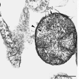
English: Fig4: Fourth to fifth day of Rickettsia helvetica infected Vero cells. a Morphology of R. helvetica in partly decomposed host cells. Note the leaflets (arrow heads) and inner plasma membrane enclosing the periplasmatic space (ps). Fibrillate nucleic acid is clearly visible (long arrows) ×120,000. Bar 150 nm. b Anti-rickettsia antibodies with gold particles (gp) (15 nm) on Lowicryl-embedded cells. Leaflets and plasma membrane are visible (arrows heads) as well as fibrillar nucleic acid (long arrows). The immunoreaction is mainly located along the membrane/leaflet part of the rickettsia but sparsely scattered all over the organism ×120,000. Bar 150 nm |
| Date | |
| Source | https://openi.nlm.nih.gov/detailedresult?img=PMC3253991_10493_2011_9508_Fig4_HTML&query=Rickettsia&it=xg&req=4&npos=10 |
| Author | Elfving K, Lukinius A, Nilsson K |
Licensing
English: This file is licensed CC BY-NC 4.0
This file was uploaded with UploadWizard.
File history
Click on a date/time to view the file as it appeared at that time.
| Date/Time | Thumbnail | Dimensions | User | Comment | |
|---|---|---|---|---|---|
| current | 21:45, 1 August 2023 | | 271 × 269 (22 KB) | Ozzie10aaaa (talk | contribs) | Uploaded a work by Elfving K, Lukinius A, Nilsson K from https://openi.nlm.nih.gov/detailedresult?img=PMC3253991_10493_2011_9508_Fig4_HTML&query=Rickettsia&it=xg&req=4&npos=10 with UploadWizard |
You cannot overwrite this file.
File usage
There are no pages that use this file.
.jpg&oldid=1253465)