File:13244 2020 892 Fig17 HTML.png
Jump to navigation
Jump to search
Size of this preview: 372 × 598 pixels. Other resolutions: 149 × 240 pixels | 298 × 480 pixels | 685 × 1,102 pixels.
Original file (685 × 1,102 pixels, file size: 334 KB, MIME type: image/png)
Summary
| Description |
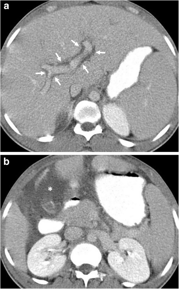
English: A 23-year-old man was admitted to the emergency department with a 1-week history of abdominal pain and diarrhea. Physical examination and blood analysis were unremarkable except for mild abdominal tenderness and eosinophilia, respectively. a, b Axial contrast-enhanced CT images demonstrate the presence of intra-abdominal free fluid (asterisk) and periportal edema (arrows). The imaging findings did not raise suspicion for an individual pathology. There was no definitive proof that this organism was the cause of the symptoms or imaging manifestation. Nevertheless, the patient’s symptoms and imaging findings were completely resolved following the treatment for Dientamoeba fragilis which was found on the patient’s stool examination |
| Date | |
| Source | https://www.ncbi.nlm.nih.gov/pmc/articles/PMC7371776/ |
| Author | Emre Ünal, Sevtap Arslan, Mehmet Ruhi Onur, and Erhan Akpinar |
Licensing
{{subst:Custom license marker added by UW}} https://creativecommons.org/licenses/by/4.0/ Attribution 4.0 International (CC BY 4.0)
This file was uploaded with UploadWizard.
File history
Click on a date/time to view the file as it appeared at that time.
| Date/Time | Thumbnail | Dimensions | User | Comment | |
|---|---|---|---|---|---|
| current | 23:31, 3 July 2023 | | 685 × 1,102 (334 KB) | Ozzie10aaaa (talk | contribs) | Uploaded a work by Emre Ünal, Sevtap Arslan, Mehmet Ruhi Onur, and Erhan Akpinar from https://www.ncbi.nlm.nih.gov/pmc/articles/PMC7371776/ with UploadWizard |
You cannot overwrite this file.
File usage
There are no pages that use this file.