File:18q syndrome (Radiopaedia 8350-9190 Axial T2 7).jpg
Jump to navigation
Jump to search
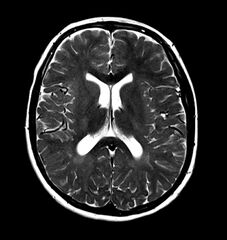
Size of this preview: 567 × 600 pixels. Other resolutions: 227 × 240 pixels | 454 × 480 pixels | 831 × 879 pixels.
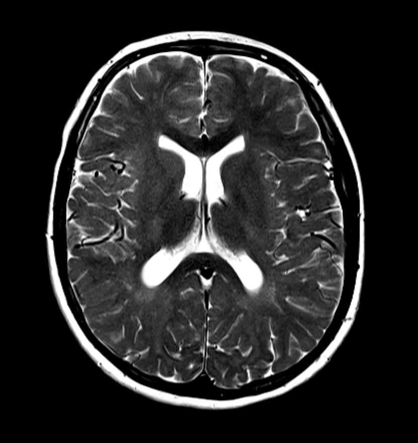
Original file (831 × 879 pixels, file size: 204 KB, MIME type: image/jpeg)
Summary:
- Radiopaedia case ID: 8350
- Image ID: 239231
- Image stack position: 7/14
- Plane projection: Axial
- Aux modality: T2
- Modality: MRI
- System: Central Nervous System
- Findings: Selected MRI images demonstrate an abnormal white matter, particularly posteriorly and in the periventricular region. It is characterized by bilateral symmetric deep white matter hyperintensity on T2 weighted images, with associated involvement of the subcortical white matter, which results in poor grey-white matter differentiation on T2 weighted images. MRS demonstrates elevation of choline (Cho). Alpha-glutamate peak is not visible. Features are consistent with subsequently established 18q syndrome.
- Published: 24th Jan 2010
- Source: https://radiopaedia.org/cases/18q-syndrome
- Author: Frank Gaillard
- Permission: http://creativecommons.org/licenses/by-nc-sa/3.0/
Licensing:
Attribution-NonCommercial-ShareAlike 3.0 Unported (CC BY-NC-SA 3.0)
File history
Click on a date/time to view the file as it appeared at that time.
| Date/Time | Thumbnail | Dimensions | User | Comment | |
|---|---|---|---|---|---|
| current | 13:20, 26 March 2021 | | 831 × 879 (204 KB) | Fæ (talk | contribs) | Radiopaedia project rID:8350 (batch #4) |
You cannot overwrite this file.
File usage
There are no pages that use this file.
.jpg&oldid=75111)