File:53662889580 26f96e4763 o.jpg
Jump to navigation
Jump to search

Size of this preview: 771 × 600 pixels. Other resolutions: 309 × 240 pixels | 617 × 480 pixels | 988 × 768 pixels | 1,280 × 995 pixels | 2,560 × 1,991 pixels | 4,123 × 3,206 pixels.
Original file (4,123 × 3,206 pixels, file size: 4.77 MB, MIME type: image/jpeg)
Summary
| Description |
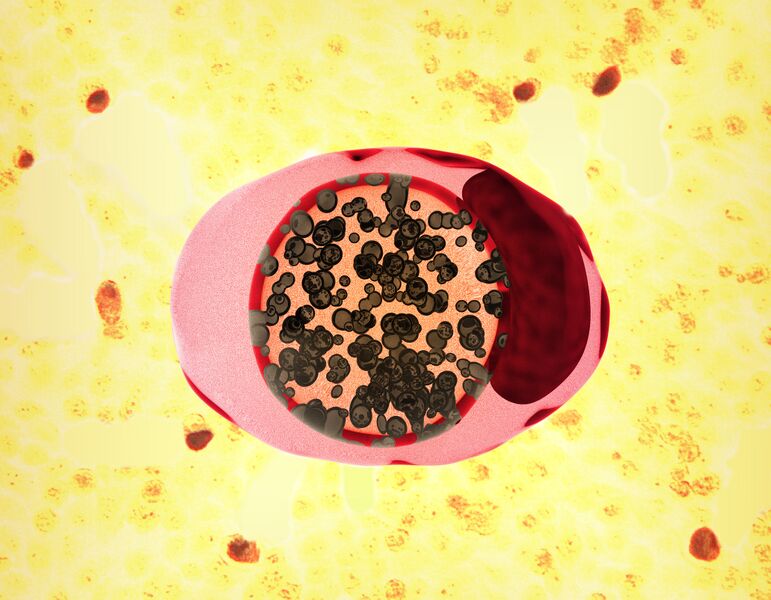
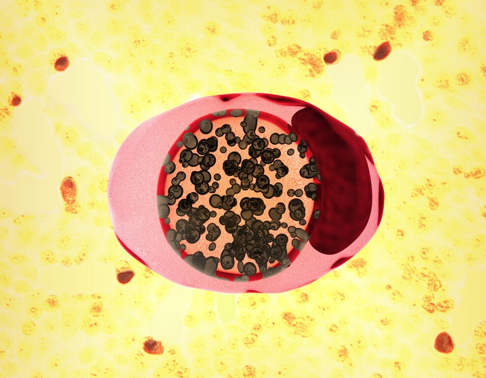
English: Illustration depicting a chlamydia-infected cell (pink and red oval shape). Chlamydia trachomatis bacteria (represented by small spherical shapes colorized brown) undergo development within a membrane-enclosed "inclusion body" (center area; orange). The dark red area on the right of the illustration denotes the cell nucleus. In the background is a CDC micrograph of cells (yellow) exhibiting chlamydia inclusion bodies (red) |
| Date | |
| Source | https://www.flickr.com/photos/niaid/53662889580/ |
| Author | NIAID and CDC |
Licensing
{{subst:Custom license marker added by UW}} https://creativecommons.org/licenses/by/2.0/ Attribution 2.0 Generic
This file was uploaded with UploadWizard.
File history
Click on a date/time to view the file as it appeared at that time.
| Date/Time | Thumbnail | Dimensions | User | Comment | |
|---|---|---|---|---|---|
| current | 22:29, 25 April 2024 | | 4,123 × 3,206 (4.77 MB) | Ozzie10aaaa (talk | contribs) | Uploaded a work by NIAID and CDC from https://www.flickr.com/photos/niaid/53662889580/ with UploadWizard |
You cannot overwrite this file.
File usage
There are no pages that use this file.