File:ACL mucoid degeration with cystic changes (Radiopaedia 48428-53341 Sagittal T1 7).jpg
Jump to navigation
Jump to search
Size of this preview: 570 × 599 pixels. Other resolutions: 228 × 240 pixels | 457 × 480 pixels | 787 × 827 pixels.
Original file (787 × 827 pixels, file size: 87 KB, MIME type: image/jpeg)
Summary:
- Radiopaedia case ID: 48428
- Image ID: 25413501
- Image stack position: 7/21
- Plane projection: Sagittal
- Aux modality: T1
- Modality: MRI
- System: Trauma
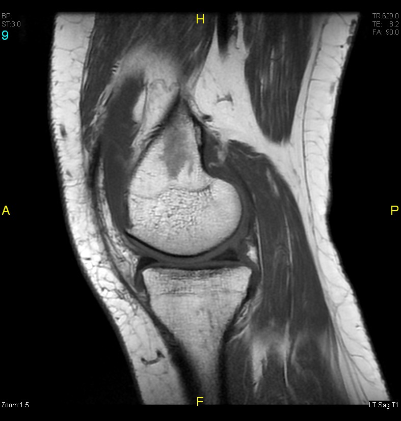
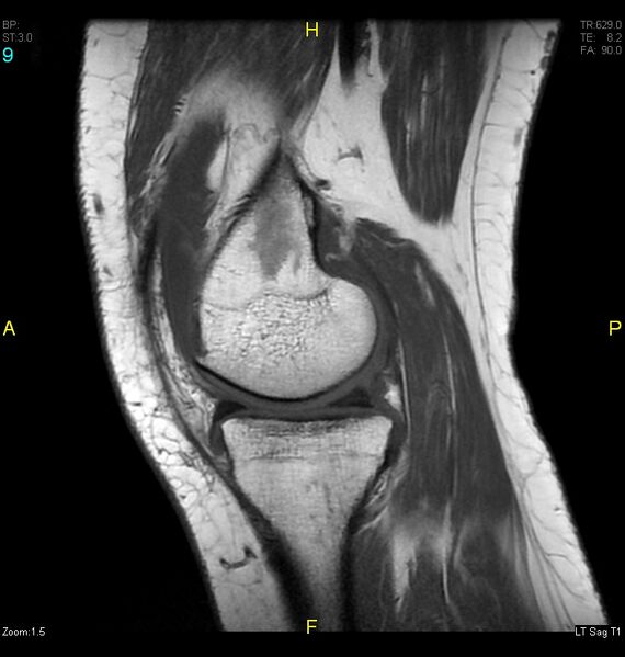
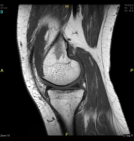
- Findings: The anterior cruciate ligament is swollen exhibiting altered intensity signal of high T2 signal and intermediate signal in T1WI. Still it showed intact bony attachment and direction of fibers, suggesting mucoid degeneration of the ACL. Small confluent cystic bony lesions are seen in subchondral part of mid upper tibia and mid posterior lower femur near the bony attachment of the ACL exhibiting hyperintense T2 signal and isointense signal in T1W. Simple cystic bony changes related to the mucoid degeneration of the ACL. Thickened medial supra-patellar plica. Marginal osteophyte formation is seen in the tibiofemoral articulation involving the margin of upper tibia, lower femur, as well as milder osteophytic changes are seen in patellofemoral relation involving the posterior and poles of patella and opposing surface. The joint space is relatively narrowed however the chondral lining is preserved. Moderate joint effusion.
- Published: 6th Oct 2016
- Source: https://radiopaedia.org/cases/acl-mucoid-degeration-with-cystic-changes
- Author: Fakhry Mahmoud Ebouda
- Permission: http://creativecommons.org/licenses/by-nc-sa/3.0/
Licensing:
Attribution-NonCommercial-ShareAlike 3.0 Unported (CC BY-NC-SA 3.0)
File history
Click on a date/time to view the file as it appeared at that time.
| Date/Time | Thumbnail | Dimensions | User | Comment | |
|---|---|---|---|---|---|
| current | 08:45, 1 April 2021 | | 787 × 827 (87 KB) | Fæ (talk | contribs) | Radiopaedia project rID:48428 (batch #459-27 B7) |
You cannot overwrite this file.
File usage
There are no pages that use this file.
.jpg&oldid=127425)