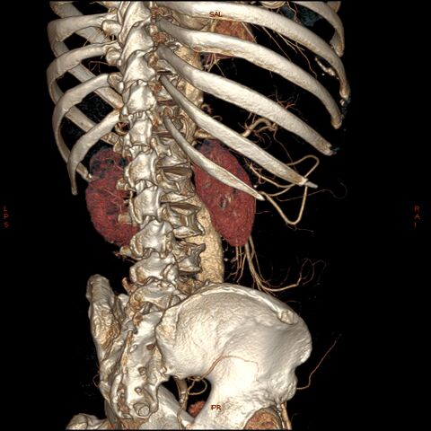
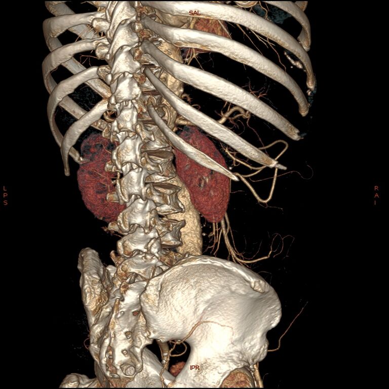
File:Abdominal aortic aneurysm (Radiopaedia 57318-64259 D 24).jpg
Jump to navigation
Jump to search

Size of this preview: 600 × 600 pixels. Other resolutions: 240 × 240 pixels | 480 × 480 pixels | 768 × 768 pixels | 1,024 × 1,024 pixels.
Original file (1,024 × 1,024 pixels, file size: 315 KB, MIME type: image/jpeg)
Summary:
- Radiopaedia case ID: 57318
- Image ID: 34680354
- Image stack position: 24/36
- Modality: CT
- System: Vascular
- Findings: 5.3 cm below the right renal artery is a fusiform dilatation of abdominal aorta with maximal cross-section diameter of 5.1 cm. Only 24% patent lumen within surrounded by chronic peripheral thrombus. The aneurysm stops short of terminal aortic bifurcation. Areas of mural calcification seen in the aorta. No features of impending rupture. Midline anterior abdominal wall hernia containing fat.
- Published: 19th Dec 2017
- Source: https://radiopaedia.org/cases/abdominal-aortic-aneurysm-30
- Author: Varun Babu
- Permission: http://creativecommons.org/licenses/by-nc-sa/3.0/
Licensing:
CC-BY-NC-SA-3.0
File history
Click on a date/time to view the file as it appeared at that time.
| Date/Time | Thumbnail | Dimensions | User | Comment | |
|---|---|---|---|---|---|
| current | 15:42, 27 March 2021 | | 1,024 × 1,024 (315 KB) | Fæ (talk | contribs) | Radiopaedia project rID:57318 (batch #75-180 D24) |
You cannot overwrite this file.
File usage
There are no pages that use this file.
.jpg&oldid=8756)