File:Abdominal aortic aneurysm (Radiopaedia 83094-97462 Sagittal renal cortical phase 126).jpg
Jump to navigation
Jump to search
Size of this preview: 445 × 599 pixels. Other resolutions: 178 × 240 pixels | 508 × 684 pixels.
Original file (508 × 684 pixels, file size: 22 KB, MIME type: image/jpeg)
Summary:
- Radiopaedia case ID: 83094
- Image ID: 53675057
- Image stack position: 126/132
- Plane projection: Sagittal
- Aux modality: renal cortical phase
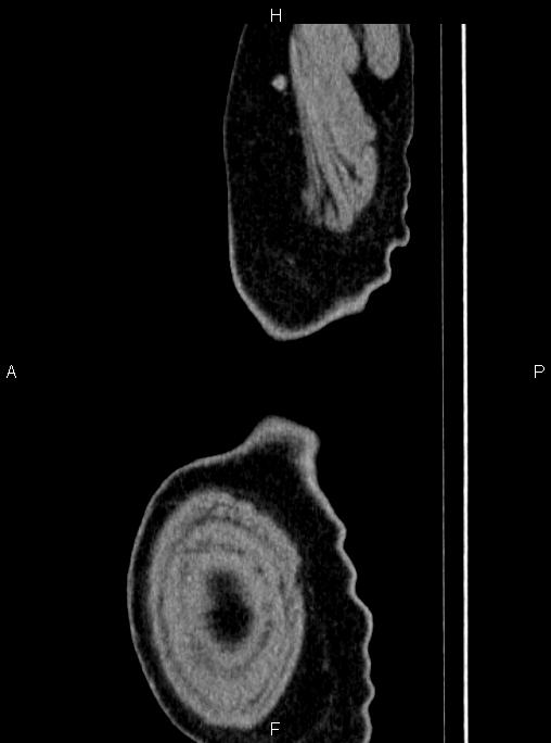
- Study findings: A few small cysts are seen at liver less than 7mm.Infrarenal fusiform abdominal aortic aneurysm is present with maximum diameter of 65mm. Marked mural thrombosis is evident up to 38mm in thickness. Contrast filled luminal caliber measured about 28mm. There is no sign of intraperitoneal rupture. Para-aortic fat is intact.
- Modality: CT
- System: Vascular
- Findings: A few small cysts are seen at liver less than 7mm. Infrarenal fusiform abdominal aortic aneurysm is present with maximum diameter of 65mm. Marked mural thrombosis is evident up to 38mm in thickness. Contrast filled luminal caliber measured about 28mm. There is no sign of intraperitoneal rupture. Para-aortic fat is intact.
- Published: 14th Oct 2020
- Source: https://radiopaedia.org/cases/abdominal-aortic-aneurysm-39
- Author: Mohammad Taghi Niknejad
- Permission: http://creativecommons.org/licenses/by-nc-sa/3.0/
Licensing:
Attribution-NonCommercial-ShareAlike 3.0 Unported (CC BY-NC-SA 3.0)
File history
Click on a date/time to view the file as it appeared at that time.
| Date/Time | Thumbnail | Dimensions | User | Comment | |
|---|---|---|---|---|---|
| current | 21:58, 26 March 2021 | | 508 × 684 (22 KB) | Fæ (talk | contribs) | Radiopaedia project rID:83094 (batch #51 D126) |
You cannot overwrite this file.
File usage
The following page uses this file:
.jpg&oldid=79497)