File:Abdominal aortic aneurysm (Radiopaedia 85063-100606 Coronal C+ arterial phase 53).jpg
Jump to navigation
Jump to search
Size of this preview: 443 × 600 pixels. Other resolutions: 177 × 240 pixels | 508 × 688 pixels.
Original file (508 × 688 pixels, file size: 57 KB, MIME type: image/jpeg)
Summary:
- Radiopaedia case ID: 85063
- Image ID: 54061183
- Image stack position: 53/58
- Plane projection: Coronal
- Aux modality: C+ arterial phase
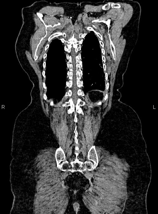
- Study findings: Post-operative changes are seen due to sternotomy and CABG.A few small nodules, less than 8mm are scattered at both lungs which seems to be fibrotic nodules. There are also several atelectatic bands scattered bilaterally. Cardiomegaly is present and ascending aorta are dilated up to 42mm. Degenerative changes as osteophytosis are seen at the thoracic spine.Large size sliding hiatal hernia is noted.Aneurysmal dilatation up to 48 mm is noted at infrarenal portion of abdominal aorta that contains mural thrombosis of 18 mm in maximum thickness. No evidence of contrast media leakage is noted. The gallbladder is not seen at anatomical location due to prior resection.A few non-enhanced simple cortical cysts are seen at both kidneys, with maximum diameters of 65mm. There are also several small parapelvic cysts at both kidneys. Fat containing small para-umbilical hernia is noted. Degenerative changes as osteophytosis are seen at the lumbar spine.
- Modality: CT
- System: Vascular
- Findings: Post-operative changes are seen due to sternotomy and CABG. A few small nodules, less than 8mm are scattered at both lungs which seems to be fibrotic nodules. There are also several atelectatic bands scattered bilaterally. Cardiomegaly is present and ascending aorta are dilated up to 42mm. Degenerative changes as osteophytosis are seen at the thoracic spine. Large size sliding hiatal hernia is noted. Aneurysmal dilatation up to 48 mm is noted at infrarenal portion of abdominal aorta that contains mural thrombosis of 18 mm in maximum thickness. No evidence of contrast media leakage is noted. The gallbladder is not seen at anatomical location due to prior resection. A few non-enhanced simple cortical cysts are seen at both kidneys, with maximum diameters of 65mm. There are also several small parapelvic cysts at both kidneys. Fat containing small para-umbilical hernia is noted. Degenerative changes as osteophytosis are seen at the lumbar spine.
- Published: 14th Dec 2020
- Source: https://radiopaedia.org/cases/abdominal-aortic-aneurysm-41
- Author: Mohammad Taghi Niknejad
- Permission: http://creativecommons.org/licenses/by-nc-sa/3.0/
Licensing:
Attribution-NonCommercial-ShareAlike 3.0 Unported (CC BY-NC-SA 3.0)
File history
Click on a date/time to view the file as it appeared at that time.
| Date/Time | Thumbnail | Dimensions | User | Comment | |
|---|---|---|---|---|---|
| current | 18:47, 26 March 2021 | | 508 × 688 (57 KB) | Fæ (talk | contribs) | Radiopaedia project rID:85063 (batch #49 E53) |
You cannot overwrite this file.
File usage
The following page uses this file:
.jpg&oldid=77674)