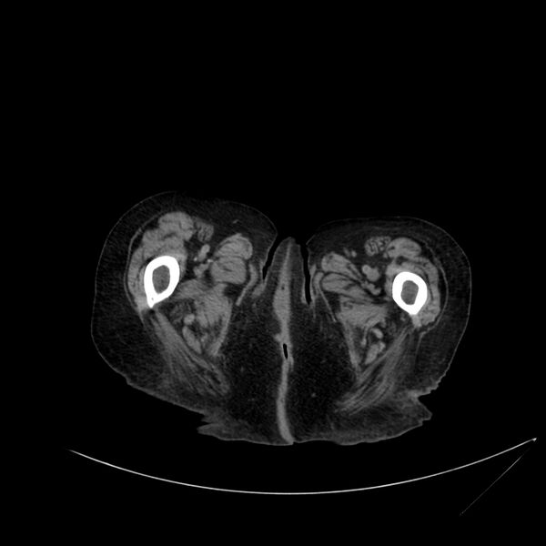
File:Abdominal aortic aneurysm - impending rupture (Radiopaedia 19233-19246 Axial non-contrast 144).jpg
Jump to navigation
Jump to search

Size of this preview: 600 × 600 pixels. Other resolutions: 240 × 240 pixels | 480 × 480 pixels | 768 × 768 pixels | 1,024 × 1,024 pixels | 1,287 × 1,287 pixels.
Original file (1,287 × 1,287 pixels, file size: 141 KB, MIME type: image/jpeg)
Summary:
- Radiopaedia case ID: 19233
- Image ID: 2280168
- Image stack position: 144/155
- Plane projection: Axial
- Aux modality: non-contrast
- Modality: CT
- System: Vascular
- Findings: CT-scan shows a complex aortic aneurysm (trilobulated abdominal aortic aneurysm with distal thoracic aneurysm) involving both the thoracic and abdominal aorta. There is an important right iliac aneurysm. In the most distal lobulation of the abdominal aneurysm, there is a hyperdense crescent associated with posterior linear fat stranding. The hyperdense crescent is easier to see on the NECT with the modified window. There is no sign of rupture. However, these findings are highly suggestive of impending rupture. There is an obstruction of the small intestine involving the jejunum at the midline. The zone of transition is located right behind the umbilicus, near a laparoscopy trocar site. The findings are compatible with a small bowel obstruction secondary to a post-operative adhesion. There is some free fluid surrounding the small bowel, but none of it has a hemorrhagic density on CT, with perihepatosplenic free fluid. Bilateral pleural effusion.
- Published: 19th Aug 2012
- Source: https://radiopaedia.org/cases/abdominal-aortic-aneurysm-impending-rupture
- Author: Maxime St-Amant
- Permission: http://creativecommons.org/licenses/by-nc-sa/3.0/
Licensing:
CC-BY-NC-SA-3.0
File history
Click on a date/time to view the file as it appeared at that time.
| Date/Time | Thumbnail | Dimensions | User | Comment | |
|---|---|---|---|---|---|
| current | 19:46, 27 March 2021 |  | 1,287 × 1,287 (141 KB) | Fæ (talk | contribs) | Radiopaedia project rID:19233 (batch #78-144 A144) |
You cannot overwrite this file.
File usage
The following page uses this file: