File:Abdominal aortic aneurysm with intramural hematoma then rupture (Radiopaedia 50278-55632 Axial C+ arterial phase 66).jpg
Jump to navigation
Jump to search

Size of this preview: 600 × 600 pixels. Other resolutions: 240 × 240 pixels | 480 × 480 pixels | 768 × 768 pixels | 1,024 × 1,024 pixels.
Original file (1,024 × 1,024 pixels, file size: 290 KB, MIME type: image/jpeg)
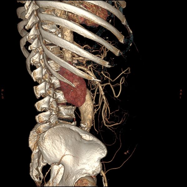
Summary:
- Radiopaedia case ID: 50278
- Image ID: 27316693
- Image stack position: 66/182
- Plane projection: Axial
- Aux modality: C+ arterial phase
- Study description: Repeat CTA 11 hours later after transfer to tertiary center
- Study findings: There is now more left retroperitoneal stranding and fluid which is displacing the left kidney anteriorly suggesting ongoing slow rupture. In the left posterolateral wall of the lower lobe of the bilobed AAA, there is contrast now extending into the wall in keeping with progression of the acute intramural hematoma. This is at the site of the wall thickening and crescent sign on the initial CT. No active contrast blush is seen outside the aorta. Again, the kidneys appear well perfused.
- Modality: CT
- System: Vascular
- Findings: The non contrast phase demonstrates a bilobed suprarenal AAA extending to the bifurcation with some subtle peripheral hyperdensity in the lower lobe forming a crescent concerning for intramural hematoma. There is some mild left retroperitoneal stranding. The CTA demonstrates eccentrically thickened aortic wall in the lower lobe of the aneurysm. No focal contrast blush is seen outside the aorta. The kidneys appear well perfused.
- Published: 12th Apr 2017
- Source: https://radiopaedia.org/cases/abdominal-aortic-aneurysm-with-intramural-haematoma-then-rupture
- Author: Craig Hacking
- Permission: http://creativecommons.org/licenses/by-nc-sa/3.0/
Licensing:
CC-BY-NC-SA-3.0
File history
Click on a date/time to view the file as it appeared at that time.
| Date/Time | Thumbnail | Dimensions | User | Comment | |
|---|---|---|---|---|---|
| current | 23:05, 27 March 2021 | | 1,024 × 1,024 (290 KB) | Fæ (talk | contribs) | Radiopaedia project rID:50278 (batch #84-66 A66) |
You cannot overwrite this file.
File usage
The following 2 files are duplicates of this file (more details):
.jpg)
.jpg)
.jpg)
The following page uses this file:
.jpg&oldid=1668452)