File:Abdominal collection due to previous cecal perforation (Radiopaedia 80831-94320 Axial C+ portal venous phase 226).jpg
Jump to navigation
Jump to search
No higher resolution available.
Abdominal_collection_due_to_previous_cecal_perforation_(Radiopaedia_80831-94320_Axial_C+_portal_venous_phase_226).jpg (512 × 512 pixels, file size: 96 KB, MIME type: image/jpeg)
Summary:
- Radiopaedia case ID: 80831
- Image ID: 53251671
- Image stack position: 226/231
- Plane projection: Axial
- Aux modality: C+ portal venous phase
- Modality: CT
- System: Gastrointestinal
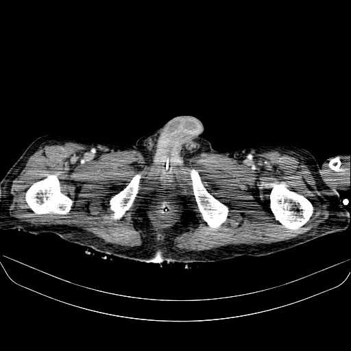
- Findings: A large cavity containing air and contrast material is seen, which extends from the anterior pelvic cavity up to the right subphrenic space, leading to bulging of the right side of the abdomen. The cavity mimics the dilated colon and may be missed easily. A focus of anterior cecal wall defect is seen that contrast passes through it into the mentioned cavity. Few fistulous tracts between the cavity and the cecum are also visible. The contrast within the bladder in the corticomedullary phase is the excreted contrast of a previous contrast-enhanced study.
- Published: 13th Aug 2020
- Source: https://radiopaedia.org/cases/abdominal-collection-due-to-previous-cecal-perforation
- Author: Faeze Salahshour
- Permission: http://creativecommons.org/licenses/by-nc-sa/3.0/
Licensing:
CC-BY-NC-SA-3.0
File history
Click on a date/time to view the file as it appeared at that time.
| Date/Time | Thumbnail | Dimensions | User | Comment | |
|---|---|---|---|---|---|
| current | 04:32, 28 March 2021 | | 512 × 512 (96 KB) | Fæ (talk | contribs) | Radiopaedia project rID:80831 (batch #95-226 A226) |
You cannot overwrite this file.
File usage
The following page uses this file:
.jpg&oldid=11794)