File:Abdominal hematoma (Radiopaedia 33614-34715 Axial C+ portal venous phase 109).png
Jump to navigation
Jump to search
No higher resolution available.
Abdominal_hematoma_(Radiopaedia_33614-34715_Axial_C+_portal_venous_phase_109).png (512 × 512 pixels, file size: 201 KB, MIME type: image/png)
Summary:
- Radiopaedia case ID: 33614
- Image ID: 10500877
- Image stack position: 109/149
- Plane projection: Axial
- Aux modality: C+ portal venous phase
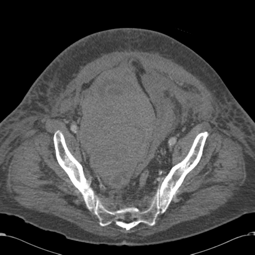
- Study findings: Right pelvic side wall hematoma, which displaces the bladder to the left. This is an extraperitoneal bleed, associated with tracking of blood in the anterior paravesical space and a small amount of stranding in the lateral posterior pararenal space. Two regions demonstrate minor arterial bleeding, in the anterosuperior and posterolateral parts, almost certainly arising from branches of the right internal iliac artery. This artery is patent proximally.
- Modality: CT
- System: Gastrointestinal
- Findings: There are a number of distended loops of small bowel present, in addition to displacement from a soft tissue mass in the pelvis. No definite evidence of free gas or obstruction.
- Published: 18th Jan 2015
- Source: https://radiopaedia.org/cases/abdominal-haematoma
- Author: RMH Core Conditions
- Permission: http://creativecommons.org/licenses/by-nc-sa/3.0/
Licensing:
CC-BY-NC-SA-3.0
File history
Click on a date/time to view the file as it appeared at that time.
| Date/Time | Thumbnail | Dimensions | User | Comment | |
|---|---|---|---|---|---|
| current | 06:25, 28 March 2021 | | 512 × 512 (201 KB) | Fæ (talk | contribs) | Radiopaedia project rID:33614 (batch #105-109 A109) |
You cannot overwrite this file.
File usage
The following page uses this file:
.png&oldid=12442)