File:Abdominal multi-trauma - devascularised kidney and liver, spleen and pancreatic lacerations (Radiopaedia 34984-36486 J 56).png
Jump to navigation
Jump to search
Size of this preview: 182 × 597 pixels. Other resolutions: 73 × 240 pixels | 512 × 1,680 pixels.
Original file (512 × 1,680 pixels, file size: 851 KB, MIME type: image/png)
Summary:
- Radiopaedia case ID: 34984
- Image ID: 11712851
- Image stack position: 56/98
- Plane projection: Sagittal
- Aux modality: bone window
- Modality: CT
- System: Hepatobiliary
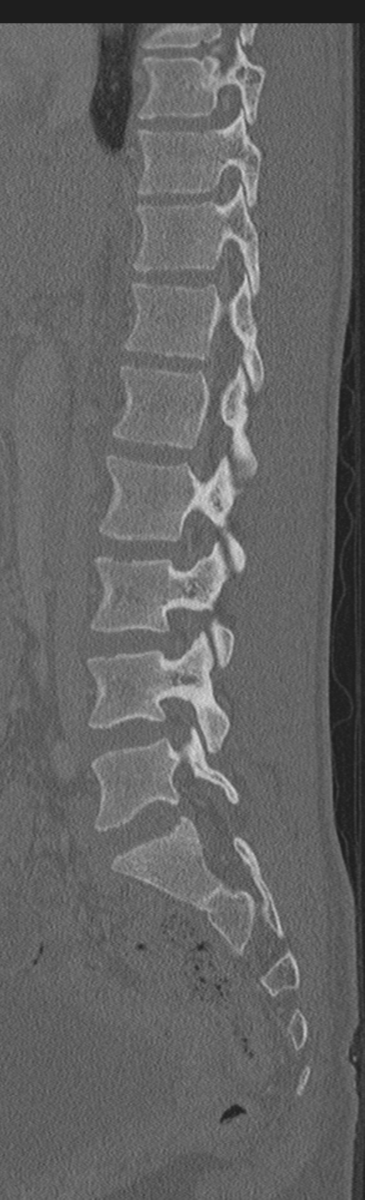
- Findings: CT Angiogram Thoracic Aorta No traumatic aortic injury or mediastinal hematoma. No pericardial effusion. There is a small right pleural collection that does not demonstrate water attenuation. There is minor bilateral dependent lung change. No focal region of consolidation. No pneumothorax. No rib or sternal fracture. CT Abdomen / Pelvis There is no perfusion of the left kidney. The left renal artery demonstrates normal opacification within its proximal 7 mm. There is however immediate truncation of the artery at a point of retroperitoneal hematoma. This hematoma lies immediately posterior to a laceration of the body of the pancreas. There are only small contrast opacified vessels identified within the left kidney. Within the posterior aspect of the left kidney, there is a region of apparent contrast extravasation that demonstrates increased spread and size increase on the delayed scan. Remote from the left renal hilum, no large left perinephric collection. On the delayed phase, there is minor contrast opacification of the left renal collecting system without evidence of injury to the left collecting system. The right kidney perfuses normally. The right renal artery has an unremarkable appearance. No evidence of traumatic injury to the right collecting system. There is a laceration through the body of the pancreas that extends completely from its anterior to posterior borders. There is also an 11 mm laceration through the medial border of the spleen that does not extend to the splenic hilum. There is associated retroperitoneal hematoma that lies within the lienorenal and gastrosplenic ligaments, as well as lying superior to the pancreas. No free intraperitoneal fluid or gas. The left adrenal gland is bulky and mildly hyperintense, likely reflecting hemorrhage. There is a laceration through the lateral segments of the left lobe of the liver. The hepatic veins, porta hepatis and intrahepatic IVC are not involved. Previous cholecystectomy. Common bile duct measures 8 mm. The ostia of the celiac trunk, SMA and IMA are unremarkable. The small and large bowel enhance normally, with no abnormal regions of abnormal wall thickening identified. Retroverted uterus. No pelvic fracture. CT Thoracic and Lumbar Spine There are fractures through the right transverse processes of T7, T8 and T9. There is a fracture through the right posterolateral superior corner of the T10 vertebral body that does not extend into the adjacent pedicle. No lumbar spine fracture. Failure of fusion of the posterior elements of S1 noted. Conclusion
- Published: 28th Aug 2015
- Source: https://radiopaedia.org/cases/abdominal-multi-trauma-devascularised-kidney-and-liver-spleen-and-pancreatic-lacerations
- Author: Craig Hacking
- Permission: http://creativecommons.org/licenses/by-nc-sa/3.0/
Licensing:
CC-BY-NC-SA-3.0
File history
Click on a date/time to view the file as it appeared at that time.
| Date/Time | Thumbnail | Dimensions | User | Comment | |
|---|---|---|---|---|---|
| current | 11:55, 28 March 2021 | 512 × 1,680 (851 KB) | Fæ (talk | contribs) | Radiopaedia project rID:34984 (batch #113-757 J56) |
You cannot overwrite this file.
File usage
The following page uses this file:
.png&oldid=14256)