File:Abdominal tuberculosis and appendicitis (Radiopaedia 65631-74735 Axial C+ portal venous phase 24).png
Jump to navigation
Jump to search
No higher resolution available.
Abdominal_tuberculosis_and_appendicitis_(Radiopaedia_65631-74735_Axial_C+_portal_venous_phase_24).png (512 × 512 pixels, file size: 171 KB, MIME type: image/png)
Summary:
- Radiopaedia case ID: 65631
- Image ID: 45590360
- Image stack position: 24/75
- Plane projection: Axial
- Aux modality: C+ portal venous phase
- Study description: CT Abdomen
- Modality: CT
- System: Gastrointestinal
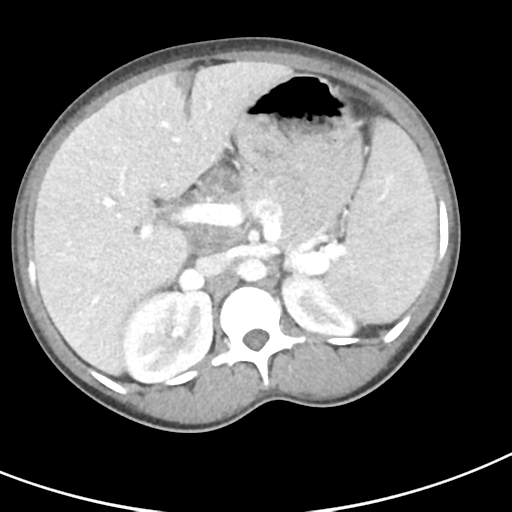
- Findings: The terminal ileum appears diffusely abnormal with dilatation, prominent wall thickening and wall enhancement. No evidence of bowel obstruction. There are multiple ring-enhancing lesions with central low attenuation measuring up to 9.2cm x 2.5 cm (axial plane) within the central mesentery extending into the right iliac fossa, suggestive of the presence of multiple necrotic lymph nodes. Mildly enlarged inguinal lymph nodes bilaterally. The appendix is dilated (measuring up to 12 mm) and lies within the right paracolic gutter with its tip abutting the inferior border of the right liver lobe. The appendix has fat stranding and demonstrates prominent wall enhancement. No appendicolith noted, nor periappendiceal collection. Conclusion: Features are of acute appendicitis.The low-density lymph nodes raise the possibility of intra-abdominal and pelvic tuberculous disease.
- Published: 24th Feb 2019
- Source: https://radiopaedia.org/cases/abdominal-tuberculosis-and-appendicitis
- Author: Sachin Phakey
- Permission: http://creativecommons.org/licenses/by-nc-sa/3.0/
Licensing:
CC-BY-NC-SA-3.0
File history
Click on a date/time to view the file as it appeared at that time.
| Date/Time | Thumbnail | Dimensions | User | Comment | |
|---|---|---|---|---|---|
| current | 13:15, 28 March 2021 | | 512 × 512 (171 KB) | Fæ (talk | contribs) | Radiopaedia project rID:65631 (batch #121-24 A24) |
You cannot overwrite this file.
File usage
The following page uses this file:
.png&oldid=14676)