File:Abdominal wall endometriosis (Radiopaedia 25723-25886 A 2).jpg
Jump to navigation
Jump to search
No higher resolution available.
Abdominal_wall_endometriosis_(Radiopaedia_25723-25886_A_2).jpg (306 × 427 pixels, file size: 33 KB, MIME type: image/jpeg)
Summary:
- Radiopaedia case ID: 25723
- Image ID: 5011311
- Image stack position: 2/4
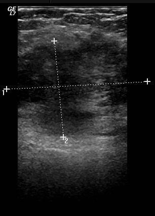
- Study findings: Ultrasonography of the lesion showed it to be ill-defined, irregular, heterogenous, hypoechoic and having some vascularity within.
- Modality: Ultrasound
- System: Gynaecology
- Findings: The lower portion of the right rectus abdominis muscle shows an irregular enhancing, mainly solid, enhancing lesion measuring 5.3 x 4.4 x 4.1 cm, related to previous cesarean section scar.
- Published: 4th Nov 2013
- Source: https://radiopaedia.org/cases/abdominal-wall-endometriosis
- Author: Ali Abougazia
- Permission: http://creativecommons.org/licenses/by-nc-sa/3.0/
Licensing:
CC-BY-NC-SA-3.0
File history
Click on a date/time to view the file as it appeared at that time.
| Date/Time | Thumbnail | Dimensions | User | Comment | |
|---|---|---|---|---|---|
| current | 15:25, 28 March 2021 | | 306 × 427 (33 KB) | Fæ (talk | contribs) | Radiopaedia project rID:25723 (batch #124-2 A2) |
You cannot overwrite this file.
File usage
There are no pages that use this file.
.jpg&oldid=15449)