File:Abdominal wall hematoma with active bleeding (Radiopaedia 83915-99122 Axial contrast-enhanced CT 29).jpg
Jump to navigation
Jump to search
No higher resolution available.
Abdominal_wall_hematoma_with_active_bleeding_(Radiopaedia_83915-99122_Axial_contrast-enhanced_CT_29).jpg (512 × 512 pixels, file size: 98 KB, MIME type: image/jpeg)
Summary:
- Radiopaedia case ID: 83915
- Image ID: 53815033
- Image stack position: 29/53
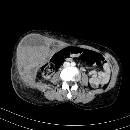
- Plane projection: Axial contrast-enhanced CT
- Study findings: The red arrows depict the foci of the active bleeding.
- Modality: Annotated image
- System: Vascular
- Findings: A large hematoma with a fluid-fluid level and another smaller similar one are visible in the right anterolateral abdominal wall. At least four foci of active bleeding are visible within the larger hematoma, which increases in size on delayed images.
- Published: 9th Nov 2020
- Source: https://radiopaedia.org/cases/abdominal-wall-haematoma-with-active-bleeding
- Author: Faeze Salahshour
- Permission: http://creativecommons.org/licenses/by-nc-sa/3.0/
Licensing:
CC-BY-NC-SA-3.0
File history
Click on a date/time to view the file as it appeared at that time.
| Date/Time | Thumbnail | Dimensions | User | Comment | |
|---|---|---|---|---|---|
| current | 18:36, 28 March 2021 | | 512 × 512 (98 KB) | Fæ (talk | contribs) | Radiopaedia project rID:83915 (batch #129-29 A29) |
You cannot overwrite this file.
File usage
The following file is a duplicate of this file (more details):
.jpg)
.jpg)
The following page uses this file:
.jpg&oldid=16102)