File:Abdominal wall metastasis from colorectal carcinoma (Radiopaedia 31016-31717 Axial C+ portal venous phase 25).jpg
Jump to navigation
Jump to search
No higher resolution available.
Abdominal_wall_metastasis_from_colorectal_carcinoma_(Radiopaedia_31016-31717_Axial_C+_portal_venous_phase_25).jpg (512 × 512 pixels, file size: 37 KB, MIME type: image/jpeg)
Summary:
- Radiopaedia case ID: 31016
- Image ID: 8303179
- Image stack position: 25/85
- Plane projection: Axial
- Aux modality: C+ portal venous phase
- Study description: Restaging CT after left external lymph node resection
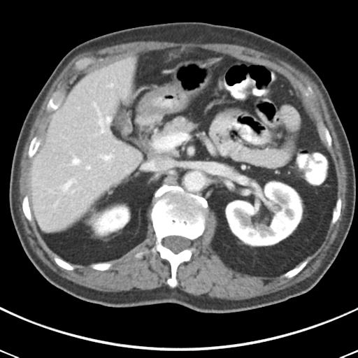
- Study findings: The previously demonstrated soft tissue mass in the left iliac fossa has beensuccessfully resected. There is no evidence of a recurrent mesenteric mass.There are surgical changes seen within the overlying left anterior abdominal wall. Inaddition there is thickening of the musculature in the left anterior abdominal wall inthis region, with the impression of a possible low density, rim enhancing lesionwithin the musculature. This has a maximum diameter of 21 mm and may represent a focalc ollection. Note is made of a surgical anastomotic line in the right colon. There is no evidenceof recurrent mass in this region.There is impression of diffuse bowel wall thickening over long segment extending fromthe rectosigmoid junction to the anal verge. This may be within normal limits althougha mass cannot be excluded. The remainder of the bowel is unremarkable. The liver, spleen, pancreas, gall bladder, both adrenals and both kidneys are grosslynormal.The prostate is markedly enlarged and heterogenous.There is moderate calcific atherosclerosis in the abdominal aorta.There is no free fluid in the abdomen and pelvis.There is no significant abdominal, pelvic or inguinal lymphadenopathy. There are degenerative changes in the lumbar spine but no sinister osseous lesions. CONCLUSION: 1. 2.1 cm rim enhancing lesion within the left lateral anterior abdominal wallmusculature. This may represent a collection or mass. Further evaluation with ultrasound is recommended. 2. Marked prostatomegaly. 3. Diffuse wall thickening throughout the rectum and rectosigmoid junction. A mass cannot be excluded and direct visualization with colonoscopy is recommended.
- Modality: CT
- System: Gastrointestinal
- Findings: FDG avid node left external iliac chain.
- Published: 20th Sep 2014
- Source: https://radiopaedia.org/cases/abdominal-wall-metastasis-from-colorectal-carcinoma
- Author: Jan Frank Gerstenmaier
- Permission: http://creativecommons.org/licenses/by-nc-sa/3.0/
Licensing:
CC-BY-NC-SA-3.0
File history
Click on a date/time to view the file as it appeared at that time.
| Date/Time | Thumbnail | Dimensions | User | Comment | |
|---|---|---|---|---|---|
| current | 19:41, 28 March 2021 | | 512 × 512 (37 KB) | Fæ (talk | contribs) | Radiopaedia project rID:31016 (batch #130-25 A25) |
You cannot overwrite this file.
File usage
The following page uses this file:
.jpg&oldid=16490)