File:Abducens nerve palsy (Radiopaedia 51069-56648 Axial DWI 12).png
Jump to navigation
Jump to search
Abducens_nerve_palsy_(Radiopaedia_51069-56648_Axial_DWI_12).png (512 × 512 pixels, file size: 83 KB, MIME type: image/png)
Summary:
- Radiopaedia case ID: 51069
- Image ID: 28172020
- Image stack position: 12/54
- Plane projection: Axial
- Aux modality: DWI
- Modality: MRI
- System: Central Nervous System
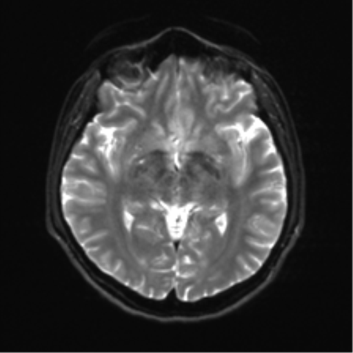
- Findings: Ovoid T2 hyperintense lesion in the posterior aspect of the pons just to the left of midline measuring 3 mm. The lesion is in the expected region of the left abducens nucleus. No associated contrast enhancement, diffusion restriction or expansion. The cisternal portion of the left abducens nerve is smaller than the right, but appears intact. Medial deviation of the left globe. The left lateral rectus demonstrates moderate atrophy. The remainder of the extra-ocular muscles have a normal appearance. Conclusion: Stable appearance of a longstanding pontine T2 hyperintense lesion in the region of the left abducens nucleus. This is most likely the cause of the left abducens nerve paresis, evidenced by medial gaze deviation and lateral rectus atrophy. Smaller left abducens nerve compared to the right presumably represents atrophy secondary to the involved abducens nucleus.
- Published: 8th Feb 2017
- Source: https://radiopaedia.org/cases/abducens-nerve-palsy
- Author: Frank Gaillard
- Permission: http://creativecommons.org/licenses/by-nc-sa/3.0/
Licensing:
CC-BY-NC-SA-3.0
File history
Click on a date/time to view the file as it appeared at that time.
| Date/Time | Thumbnail | Dimensions | User | Comment | |
|---|---|---|---|---|---|
| current | 21:59, 28 March 2021 |  | 512 × 512 (83 KB) | Fæ (talk | contribs) | Radiopaedia project rID:51069 (batch #136-82 E12) |
You cannot overwrite this file.
File usage
The following page uses this file:
