File:Aberrant right subclavian and left vertebral arteries (Radiopaedia 43467-46847 Axial C+ portal venous phase 71).jpg
Jump to navigation
Jump to search

Size of this preview: 600 × 600 pixels. Other resolutions: 240 × 240 pixels | 480 × 480 pixels | 768 × 768 pixels | 1,024 × 1,024 pixels | 2,048 × 2,048 pixels | 4,004 × 4,004 pixels.
Original file (4,004 × 4,004 pixels, file size: 2.07 MB, MIME type: image/jpeg)
Summary:
- Radiopaedia case ID: 43467
- Image ID: 20160829
- Image stack position: 71/76
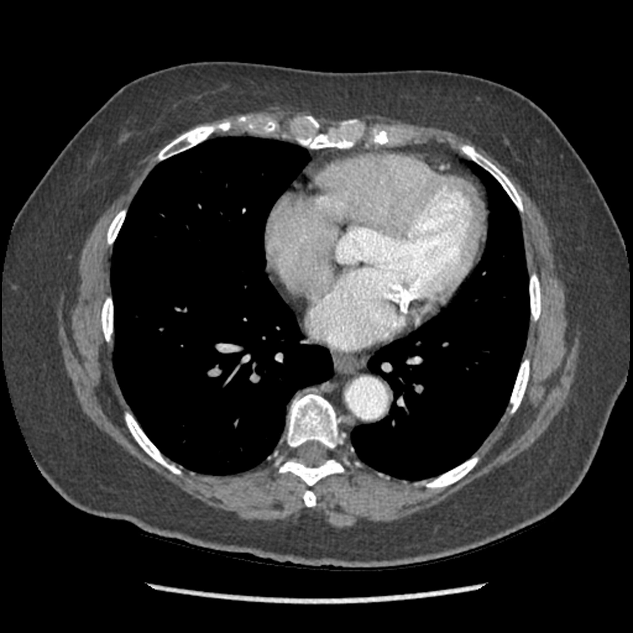
- Plane projection: Axial
- Aux modality: C+ portal venous phase
- Modality: CT
- System: Chest
- Findings: Contrast enhanced CT demonstrates an aberrant right subclavian artery, and aberrant origin of the left vertebral artery proximal to origin of the left subclavian artery. The patient is status post right thyroidectomy. Gallstones seen in the upper abdomen scan.
- Published: 11th Mar 2017
- Source: https://radiopaedia.org/cases/aberrant-right-subclavian-and-left-vertebral-arteries
- Author: Hani Makky Al Salam
- Permission: http://creativecommons.org/licenses/by-nc-sa/3.0/
Licensing:
CC-BY-NC-SA-3.0
File history
Click on a date/time to view the file as it appeared at that time.
| Date/Time | Thumbnail | Dimensions | User | Comment | |
|---|---|---|---|---|---|
| current | 11:06, 29 March 2021 | | 4,004 × 4,004 (2.07 MB) | Fæ (talk | contribs) | Radiopaedia project rID:43467 (batch #146-71 A71) |
You cannot overwrite this file.
File usage
The following page uses this file:
.jpg&oldid=18976)