File:Aberrant right subclavian artery (Radiopaedia 34993-36495 Coronal T2 1).jpeg
Jump to navigation
Jump to search
Size of this preview: 573 × 600 pixels. Other resolutions: 229 × 240 pixels | 459 × 480 pixels | 644 × 674 pixels.
Original file (644 × 674 pixels, file size: 108 KB, MIME type: image/jpeg)
Summary:
- Radiopaedia case ID: 34993
- Image ID: 11713346
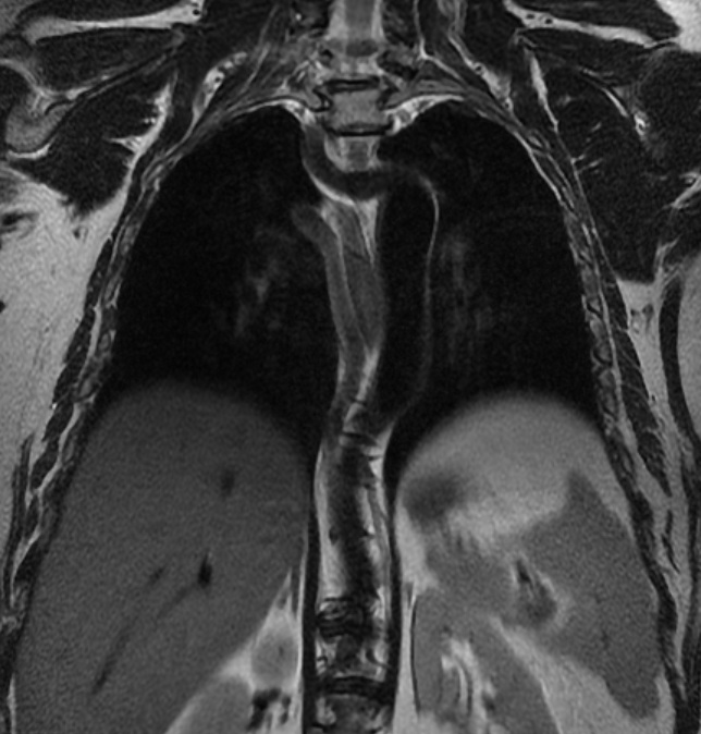
- Plane projection: Coronal
- Aux modality: T2
- Study description: Dorsal Spine
- Modality: MRI
- System: Vascular
- Findings: Hypointense rounded structure both on T1 and T2, just anterior to the dorsal spine impressing the esophagus that does not look proximally. On the coronal image, an aberrant artery originating from the aortic arch is seen.
- Published: 18th Mar 2015
- Source: https://radiopaedia.org/cases/aberrant-right-subclavian-artery-31
- Author: Enrico Citarella
- Permission: http://creativecommons.org/licenses/by-nc-sa/3.0/
Licensing:
CC-BY-NC-SA-3.0
File history
Click on a date/time to view the file as it appeared at that time.
| Date/Time | Thumbnail | Dimensions | User | Comment | |
|---|---|---|---|---|---|
| current | 13:36, 29 March 2021 | | 644 × 674 (108 KB) | Fæ (talk | contribs) | Radiopaedia project rID:34993 (batch #150-3 C1) |
You cannot overwrite this file.
File usage
There are no pages that use this file.
.jpeg&oldid=19679)