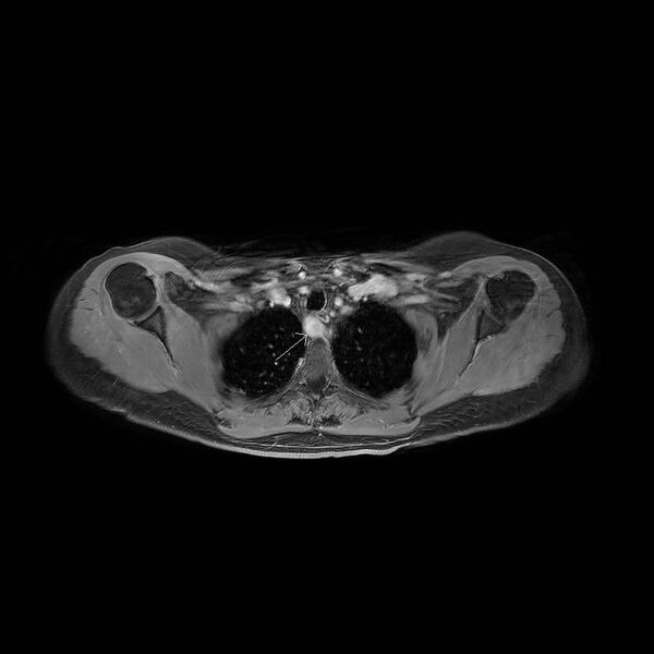
File:Aberrant right subclavian artery with background Takayasu arteritis (Radiopaedia 21423-21363 Axial MRA 16).jpg
Jump to navigation
Jump to search

Size of this preview: 600 × 600 pixels. Other resolutions: 240 × 240 pixels | 480 × 480 pixels | 768 × 768 pixels.
Original file (768 × 768 pixels, file size: 48 KB, MIME type: image/jpeg)
Summary:
- Radiopaedia case ID: 21423
- Image ID: 2876282
- Image stack position: 16/60
- Plane projection: Axial
- Aux modality: MRA
- Modality: MRI
- System: Vascular
- Findings: MR angiogram with gadolinium demonstrates an aberrant right subclavian artery (arrowed). Note normal loss of signal (flow void) due to blood changing direction within the descending aorta. Also, note the associated aneurysmal dilatation of this vessel. Coronal MR angiogram further demonstrates the aberrant origin of the right subclavian artery. Incidental gallstone noted (arrowed). 3D rendered image demonstrates the aberrant subclavian anatomy. In addition, there is considerable pruning and stenoses with areas of occlusion involving the subclavian arteries more distally (arrowed).
- Published: 22nd Jan 2013
- Source: https://radiopaedia.org/cases/aberrant-right-subclavian-artery-with-background-takayasu-arteritis
- Author: Ashok Kumar
- Permission: http://creativecommons.org/licenses/by-nc-sa/3.0/
Licensing:
CC-BY-NC-SA-3.0
File history
Click on a date/time to view the file as it appeared at that time.
| Date/Time | Thumbnail | Dimensions | User | Comment | |
|---|---|---|---|---|---|
| current | 18:20, 29 March 2021 |  | 768 × 768 (48 KB) | Fæ (talk | contribs) | Radiopaedia project rID:21423 (batch #178-16 A16) |
You cannot overwrite this file.
File usage
The following page uses this file: