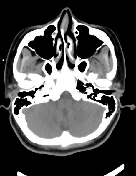
File:Accessory parotid glands (Radiopaedia 27289-27472 Axial non-contrast 20).jpg
Jump to navigation
Jump to search

Size of this preview: 462 × 599 pixels. Other resolutions: 185 × 240 pixels | 512 × 664 pixels.
Original file (512 × 664 pixels, file size: 31 KB, MIME type: image/jpeg)
Summary:
- Radiopaedia case ID: 27289
- Image ID: 5721169
- Image stack position: 20/20
- Plane projection: Axial
- Aux modality: non-contrast
- Modality: CT
- System: Head & Neck
- Findings: The right accessory parotid gland is seen lateral to the right masseter muscle, distinctly separate from the main parotid gland, with the same CT density of both parotid glands. The smaller left accessory parotid gland is seen opposite the anterior lateral aspect of the left masseter muscle.
- Published: 27th Jan 2014
- Source: https://radiopaedia.org/cases/accessory-parotid-glands
- Author: Dalia Ibrahim
- Permission: http://creativecommons.org/licenses/by-nc-sa/3.0/
Licensing:
CC-BY-NC-SA-3.0
File history
Click on a date/time to view the file as it appeared at that time.
| Date/Time | Thumbnail | Dimensions | User | Comment | |
|---|---|---|---|---|---|
| current | 13:24, 30 March 2021 |  | 512 × 664 (31 KB) | Fæ (talk | contribs) | Radiopaedia project rID:27289 (batch #268-20 A20) |
You cannot overwrite this file.
File usage
There are no pages that use this file.