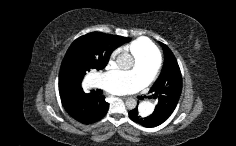
File:Accessory right inferior hepatic vein (Radiopaedia 65245-74259 Axial C+ arterial phase 21).jpg
Jump to navigation
Jump to search

Size of this preview: 800 × 494 pixels. Other resolutions: 320 × 198 pixels | 640 × 396 pixels | 1,021 × 631 pixels.
Original file (1,021 × 631 pixels, file size: 192 KB, MIME type: image/jpeg)
Summary:
- Radiopaedia case ID: 65245
- Image ID: 45120507
- Image stack position: 21/92
- Plane projection: Axial
- Aux modality: C+ arterial phase
- Modality: CT
- System: Chest
- Findings: Marked dilatation of the pulmonary trunk (6.7 cm) with the right (5.4 cm) and left (4 cm) main branches. Lung window shows mild bilateral diffuse faint groundglass centrilobular lung nodules that may reflect an underlying infection. Scans through the upper abdomen revealed average size cirrhotic liver and reflux of contrast into the IVC and hepatic veins with Incidental opacification of accessory right inferior hepatic vein. Also noted, infrarenal duplicated Inferior vena cava with azygos continuation draining at the superior vena cava. The left renal vein continues upwards replacing the hepatic segment of IVC draining the hepatic veins and the accessory right inferior hepatic vein till the right atrium. Unfortunately, the venous phase is not available and wasn't performed.
- Published: 17th Jan 2019
- Source: https://radiopaedia.org/cases/accessory-right-inferior-hepatic-vein-3
- Author: Mostafa El-Feky
- Permission: http://creativecommons.org/licenses/by-nc-sa/3.0/
Licensing:
CC-BY-NC-SA-3.0
File history
Click on a date/time to view the file as it appeared at that time.
| Date/Time | Thumbnail | Dimensions | User | Comment | |
|---|---|---|---|---|---|
| current | 14:07, 30 March 2021 |  | 1,021 × 631 (192 KB) | Fæ (talk | contribs) | Radiopaedia project rID:65245 (batch #276-21 A21) |
You cannot overwrite this file.
File usage
The following page uses this file: