File:Accessory soleus muscle (Radiopaedia 61069-68944 A 1).jpg
Jump to navigation
Jump to search

Size of this preview: 800 × 400 pixels. Other resolutions: 320 × 160 pixels | 640 × 320 pixels | 1,260 × 630 pixels.
Original file (1,260 × 630 pixels, file size: 64 KB, MIME type: image/jpeg)
Summary:
- Radiopaedia case ID: 61069
- Image ID: 40066039
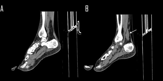
- Study findings: The white arrow refers to filling of the Kager fat pad by an accessory soleus muscle.
- Modality: Annotated image
- System: Musculoskeletal
- Findings: An accessory soleus muscle is seen at the left side. Its origin has no bony attachment and mostly from the anterior surface of the soleus muscle. Its insertion distally through a short tendon into the upper calcaneal surface.
- Published: 15th Jun 2018
- Source: https://radiopaedia.org/cases/accessory-soleus-muscle
- Author: Ahmed Abdrabou
- Permission: http://creativecommons.org/licenses/by-nc-sa/3.0/
Licensing:
CC-BY-NC-SA-3.0
File history
Click on a date/time to view the file as it appeared at that time.
| Date/Time | Thumbnail | Dimensions | User | Comment | |
|---|---|---|---|---|---|
| current | 16:15, 30 March 2021 | | 1,260 × 630 (64 KB) | Fæ (talk | contribs) | Radiopaedia project rID:61069 (batch #283-1 A1) |
You cannot overwrite this file.
File usage
There are no pages that use this file.
.jpg&oldid=28100)