File:Accessory soleus muscle (Radiopaedia 67929).jpg
Jump to navigation
Jump to search

Size of this preview: 800 × 359 pixels. Other resolutions: 320 × 144 pixels | 853 × 383 pixels.
Original file (853 × 383 pixels, file size: 71 KB, MIME type: image/jpeg)
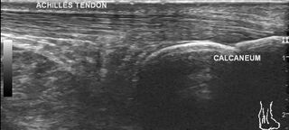
Summary:
- Radiopaedia case ID: 67929
- Image ID: 48523708
- Modality: Ultrasound
- System: Musculoskeletal
- Findings: The diffuse bulge pointed by the patient is a muscle with normal echopattern; located between Achilles tendon and flexure hallucis longus tendon and replaces the pre-Achilles fat pad. It shows continuity with the soleus muscle cranially. There is an aponeurosis in posterior most part of the above-mentioned muscle which forms a very short tendon which is inserted into the upper border of the calcaneum, medial to the Achilles tendon insertion. The Achilles tendon is normal. Flexure hallucis longus tendon is normal. Adjacent neurovascular bundle is normal. Comparison with the asymptomatic side does not show accessory muscle.
- Published: 4th May 2019
- Source: https://radiopaedia.org/cases/accessory-soleus-muscle-2
- Author: Maulik S Patel
- Permission: http://creativecommons.org/licenses/by-nc-sa/3.0/
Licensing:
CC-BY-NC-SA-3.0
File history
Click on a date/time to view the file as it appeared at that time.
| Date/Time | Thumbnail | Dimensions | User | Comment | |
|---|---|---|---|---|---|
| current | 15:06, 30 March 2021 | | 853 × 383 (71 KB) | Fæ (talk | contribs) | Radiopaedia project rID:67929 (batch #281) |
You cannot overwrite this file.
File usage
The following page uses this file:
.jpg&oldid=8860028)