File:Achalasia (Radiopaedia 14946-14878 Axial lung window 7).jpg
Jump to navigation
Jump to search

Size of this preview: 600 × 600 pixels. Other resolutions: 240 × 240 pixels | 480 × 480 pixels | 647 × 647 pixels.
Original file (647 × 647 pixels, file size: 154 KB, MIME type: image/jpeg)
Summary:
- Radiopaedia case ID: 14946
- Image ID: 1365342
- Image stack position: 7/75
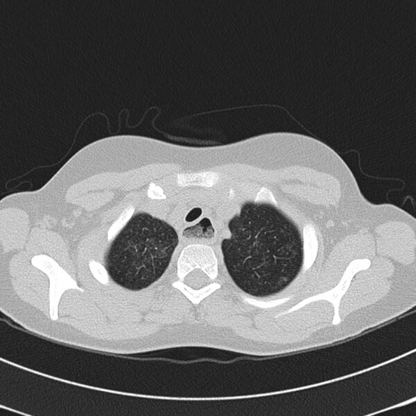
- Plane projection: Axial
- Aux modality: lung window
- Study findings: Grossly distended esophagus contains a large amount of food debris. There are numerous small foci of airspace and groundglass opacification. Given the appearance of the esophagus, the lung changes most likely reflect aspiration.
- Modality: CT
- System: Gastrointestinal
- Findings: There are prominent perihilar interstitial markings and patchy airspace opacification.
- Published: 7th Sep 2011
- Source: https://radiopaedia.org/cases/achalasia-7
- Author: Alexandra Stanislavsky
- Permission: http://creativecommons.org/licenses/by-nc-sa/3.0/
Licensing:
Attribution-NonCommercial-ShareAlike 3.0 Unported (CC BY-NC-SA 3.0)
File history
Click on a date/time to view the file as it appeared at that time.
| Date/Time | Thumbnail | Dimensions | User | Comment | |
|---|---|---|---|---|---|
| current | 21:08, 30 March 2021 | | 647 × 647 (154 KB) | Fæ (talk | contribs) | Radiopaedia project rID:14946 (batch #341-7 A7) |
You cannot overwrite this file.
File usage
The following page uses this file:
.jpg&oldid=137031)