File:Achalasia (Radiopaedia 24937-25206 Axial lung window 30).jpg
Jump to navigation
Jump to search
Size of this preview: 600 × 600 pixels. Other resolutions: 240 × 240 pixels | 631 × 631 pixels.
Original file (631 × 631 pixels, file size: 95 KB, MIME type: image/jpeg)
Summary:
- Radiopaedia case ID: 24937
- Image ID: 4721542
- Image stack position: 30/32
- Plane projection: Axial
- Aux modality: lung window
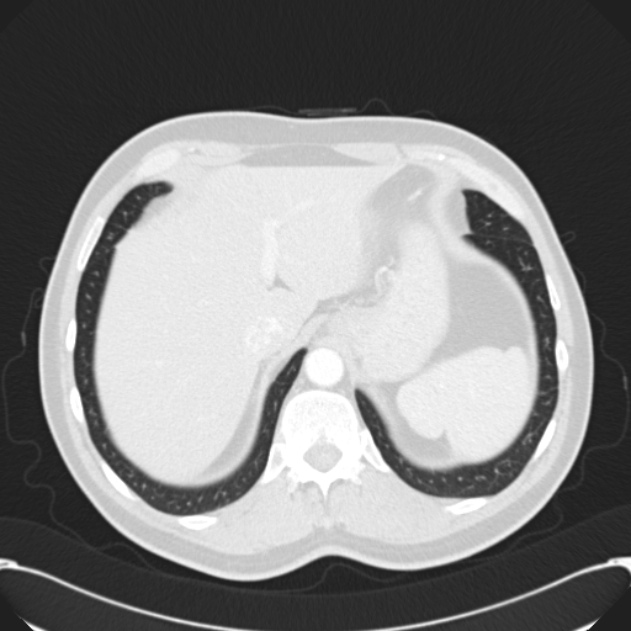
- Study findings: The esophagus is dilated and distally there is smooth circumferential wall thickening. Fluid-fluid level is present in the esophagus. There is no evidence of aspiration at this time. No lymphadenopathy.
- Modality: CT
- System: Chest
- Findings: The gastric gas bubble is absent. Lungs appear normal. Heart and mediastinal contours appear normal.
- Published: 21st Sep 2013
- Source: https://radiopaedia.org/cases/achalasia-11
- Author: Jan Frank Gerstenmaier
- Permission: http://creativecommons.org/licenses/by-nc-sa/3.0/
Licensing:
Attribution-NonCommercial-ShareAlike 3.0 Unported (CC BY-NC-SA 3.0)
File history
Click on a date/time to view the file as it appeared at that time.
| Date/Time | Thumbnail | Dimensions | User | Comment | |
|---|---|---|---|---|---|
| current | 21:57, 30 March 2021 | | 631 × 631 (95 KB) | Fæ (talk | contribs) | Radiopaedia project rID:24937 (batch #343-62 B30) |
You cannot overwrite this file.
File usage
There are no pages that use this file.
.jpg&oldid=137789)