File:Achalasia (Radiopaedia 47147-51685 Axial lung window 20).png
Jump to navigation
Jump to search
No higher resolution available.
Achalasia_(Radiopaedia_47147-51685_Axial_lung_window_20).png (512 × 512 pixels, file size: 137 KB, MIME type: image/png)
Summary:
- Radiopaedia case ID: 47147
- Image ID: 24348829
- Image stack position: 20/51
- Plane projection: Axial
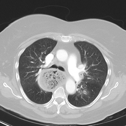
- Aux modality: lung window
- Study findings: Chest: there is severe dilatation of the esophagus extending from the thoracic inlet down to the gastro-esophageal junction. The lumen is loaded with debris. Within the limits of CT no definite obstructing mass is seen at the gastro-esophageal junction (also there is no evidence of previous surgery to indicate there has been previous esophageal interposition or Ivor Lewis procedure). No evidence of axillary or supraclavicular lymphadenopathy. Enhancement of the aortic arch and pulmonary tree are within normal limits. No evidence of a mediastinal hematoma or pericardial effusion. There are multiple mildly enlarged left anterior mediastinal and subcarinal lymph nodes. There are enlarged hilar lymph nodes bilaterally. There is diffuse airways wall thickening with peribronchovascular inflammatory changes, most marked in the left lower lobe. There are several nodular opacities, the largest in the anteroinferior aspect of the right upper lobe measuring 13 x 9 mm. There is retrocrural lymphadenopathy. Upper abdomen: the liver, spleen, kidneys, adrenals, visualized bowel and appendix are within normal limits. There is atrophic changes of the pancreas. There are calcifications within the wall of the gallbladder. No free intraperitoneal fluid or gas. The abdominal aorta is nonaneurysmal. There are at least four enlarged and centrally necrotic retroperitoneal lymph nodes, the largest measuring 24 x 20 mm inferior to the left renal vein. No destructive bony lesion identified. CONCLUSION: Gastroenterology review recommended:
- Modality: CT
- System: Gastrointestinal
- Findings: Chest x-ray: PA and lateral projections. Widening of the right superior mediastinum with mottled opacity is consistent with a dilated esophagus. 11 mm nodular opacity within the right mid-zone. Minor airspace opacity within the left medial lower zone may represent infective consolidation or aspiration. No pleural effusion. No destructive osseous lesion.
- Published: 2nd Aug 2016
- Source: https://radiopaedia.org/cases/achalasia-20
- Author: Melbourne Uni Radiology Masters
- Permission: http://creativecommons.org/licenses/by-nc-sa/3.0/
Licensing:
Attribution-NonCommercial-ShareAlike 3.0 Unported (CC BY-NC-SA 3.0)
File history
Click on a date/time to view the file as it appeared at that time.
| Date/Time | Thumbnail | Dimensions | User | Comment | |
|---|---|---|---|---|---|
| current | 00:26, 31 March 2021 | | 512 × 512 (137 KB) | Fæ (talk | contribs) | Radiopaedia project rID:47147 (batch #349-71 B20) |
You cannot overwrite this file.
File usage
The following page uses this file:
.png&oldid=138766)