File:Achalasia (Radiopaedia 8831-9629 Axial lung window 1).jpg
Jump to navigation
Jump to search
No higher resolution available.
Achalasia_(Radiopaedia_8831-9629_Axial_lung_window_1).jpg (512 × 512 pixels, file size: 136 KB, MIME type: image/jpeg)
Summary:
- Radiopaedia case ID: 8831
- Image ID: 344213
- Image stack position: 1/66
- Plane projection: Axial
- Aux modality: lung window
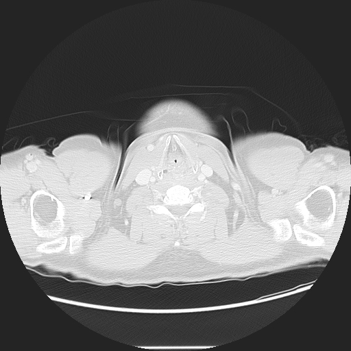
- Study description: CT chest / abdomen / pelvis
- Study findings: CT scan of the abdomen showed uniform dilatation of the esophagus with air-fluid level. Patchy consolidation in the upper segment of the right lower lobe likely due to aspiration.
- Modality: CT
- System: Gastrointestinal
- Findings: Chest x-ray is largely unremarkable. Minor patchy opacities in the left base are noted. A small gastric air bubble is visible.
- Published: 2nd Mar 2010
- Source: https://radiopaedia.org/cases/achalasia-1
- Author: Hani Makky Al Salam
- Permission: http://creativecommons.org/licenses/by-nc-sa/3.0/
Licensing:
Attribution-NonCommercial-ShareAlike 3.0 Unported (CC BY-NC-SA 3.0)
File history
Click on a date/time to view the file as it appeared at that time.
| Date/Time | Thumbnail | Dimensions | User | Comment | |
|---|---|---|---|---|---|
| current | 20:50, 30 March 2021 | | 512 × 512 (136 KB) | Fæ (talk | contribs) | Radiopaedia project rID:8831 (batch #338-54 B1) |
You cannot overwrite this file.
File usage
The following page uses this file:
.jpg&oldid=137180)