File:Achalasia of the cardia (Radiopaedia 38497-40595 Axial C+ arterial phase 1).png
Jump to navigation
Jump to search
No higher resolution available.
Achalasia_of_the_cardia_(Radiopaedia_38497-40595_Axial_C+_arterial_phase_1).png (512 × 512 pixels, file size: 137 KB, MIME type: image/png)
Summary:
- Radiopaedia case ID: 38497
- Image ID: 14711142
- Image stack position: 1/53
- Plane projection: Axial
- Aux modality: C+ arterial phase
- Study description: CT Chest and CT Abdomen
- Modality: CT
- System: Gastrointestinal
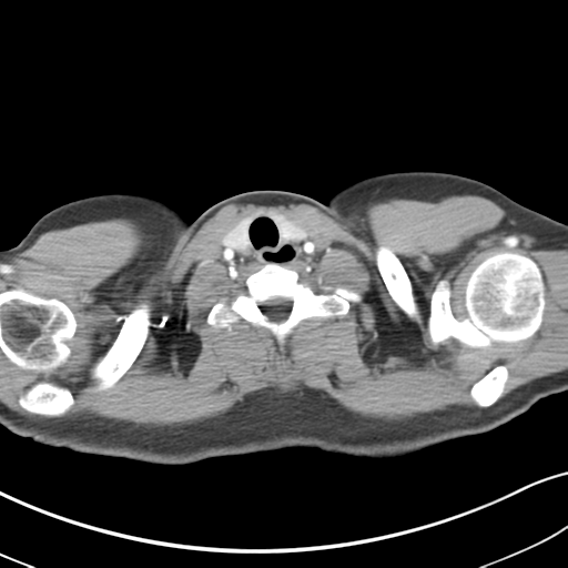
- Findings: CT chest, abdomen with Oral and IV contrast: Gross distension of the entire esophagus down to be the gastroesophageal junction, consistent with achalasia. No obvious evidence of complicating esophageal carcinoma. No mediastinal lymphadenopathy. Lung fields are unremarkable with no evidence of chronic aspiration. In the abdomen the appearances are unremarkable.
- Published: 30th Aug 2015
- Source: https://radiopaedia.org/cases/achalasia-of-the-cardia
- Author: Melbourne Uni Radiology Masters
- Permission: http://creativecommons.org/licenses/by-nc-sa/3.0/
Licensing:
Attribution-NonCommercial-ShareAlike 3.0 Unported (CC BY-NC-SA 3.0)
File history
Click on a date/time to view the file as it appeared at that time.
| Date/Time | Thumbnail | Dimensions | User | Comment | |
|---|---|---|---|---|---|
| current | 09:18, 31 March 2021 | | 512 × 512 (137 KB) | Fæ (talk | contribs) | Radiopaedia project rID:38497 (batch #357-98 C1) |
You cannot overwrite this file.
File usage
The following page uses this file:
.png&oldid=142105)