File:Achilles tendon tear (Radiopaedia 18551-18421 C 1).jpg
Jump to navigation
Jump to search
No higher resolution available.
Achilles_tendon_tear_(Radiopaedia_18551-18421_C_1).jpg (708 × 502 pixels, file size: 63 KB, MIME type: image/jpeg)
Summary:
- Radiopaedia case ID: 18551
- Image ID: 2122432
- Plane projection: Longitudinal
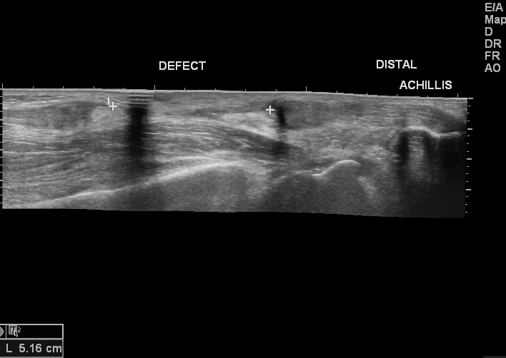
- Study findings: There is a complete tear of Achilles tendon approximately 6 cm away from the insertion site. The gap between the two edges of the tendon is 5 cm with a neutral position of the foot ( without plantar / dorsiflexion ). The torn ends of the tendon show thickening. The distal tendon near the insertion shows a normal echopattern.
- Modality: Ultrasound
- System: Trauma
- Findings: There is a presence of calcaneal spurs. Achilles tendon shadow shows thinning in the distal leg. The tendon shadow shows thickening near the insertion. Kager fat pad is partially obliterated. There is no fracture/ dislocation.
- Published: 12th Jul 2012
- Source: https://radiopaedia.org/cases/achilles-tendon-tear
- Author: Maulik S Patel
- Permission: http://creativecommons.org/licenses/by-nc-sa/3.0/
Licensing:
CC-BY-NC-SA-3.0
File history
Click on a date/time to view the file as it appeared at that time.
| Date/Time | Thumbnail | Dimensions | User | Comment | |
|---|---|---|---|---|---|
| current | 17:19, 31 March 2021 | | 708 × 502 (63 KB) | Fæ (talk | contribs) | Radiopaedia project rID:18551 (batch #404-3 C1) |
You cannot overwrite this file.
File usage
There are no pages that use this file.
.jpg&oldid=34327)