File:Achilles tendon tear (Radiopaedia 9759-10363 Lateral 1).jpg
Jump to navigation
Jump to search
Size of this preview: 374 × 600 pixels. Other resolutions: 149 × 240 pixels | 469 × 752 pixels.
Original file (469 × 752 pixels, file size: 53 KB, MIME type: image/jpeg)
Summary:
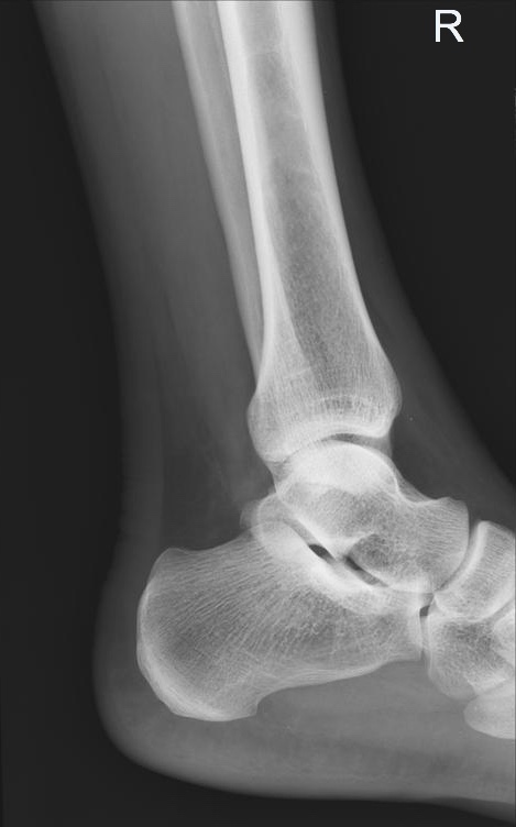
- Radiopaedia case ID: 9759
- Image ID: 52527740
- Plane projection: Lateral
- Study findings: On the lateral comparison view, the asymptomatic right Achilles tendon shows a homogeneous density and sharp margins. The symptomatic left tendon shows focal thickening and irregular contour due to the tendon tear with retracted edges. The dorsal fat pad (Kager triangle) is not opacified in the absence of fluid due to chronic injury.
- Modality: X-ray
- System: Trauma
- Findings: The asymptomatic right Achilles tendon is intact with a normal echo pattern. The symptomatic left Achilles tendon shows a complete tear with a gap between two edges measuring more than 30 mm. The distal tendon shows thickening and hypoechoic echo pattern favoring tendinosis.
- Published: 21st May 2010
- Source: https://radiopaedia.org/cases/achilles-tendon-tear-2
- Author: Maulik S Patel
- Permission: http://creativecommons.org/licenses/by-nc-sa/3.0/
Licensing:
CC-BY-NC-SA-3.0
File history
Click on a date/time to view the file as it appeared at that time.
| Date/Time | Thumbnail | Dimensions | User | Comment | |
|---|---|---|---|---|---|
| current | 17:21, 31 March 2021 | | 469 × 752 (53 KB) | Fæ (talk | contribs) | Radiopaedia project rID:9759 (batch #406-1 A1) |
You cannot overwrite this file.
File usage
There are no pages that use this file.
.jpg&oldid=34339)