File:Acoustic neuroma (Radiopaedia 34049-35283 Axial 50).png
Jump to navigation
Jump to search
No higher resolution available.
Acoustic_neuroma_(Radiopaedia_34049-35283_Axial_50).png (512 × 512 pixels, file size: 204 KB, MIME type: image/png)
Summary:
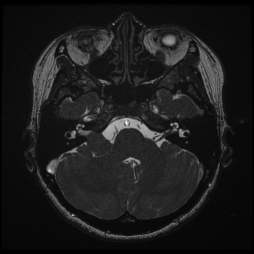
- Radiopaedia case ID: 34049
- Image ID: 10842592
- Image stack position: 50/84
- Plane projection: Axial
- Modality: MRI
- System: Oncology
- Findings: Right cerebellopontine angle mass is seen to extend into the right internal acoustic canal which it enlarges. It demonstrates mass effect and slight distortion on the right middle cerebellar peduncle but no evidence of abnormal T2/FLAIR signal to suggest edema or gliosis. The mass is characterized by low T1 signal, intermediate T2 signal with areas of cystic change and demonstrates heterogeneous enhancement. Small right Meckels cave enhancing lesion likely represents a small right trigeminal nerve schwannoma and measures 6 x 5 x 7 mm (AP x RL x CC). The left internal acoustic canal is normal with no suggestion of mass. Conclusion: 1. Right cerebellopontine angle mass demonstrates features characteristic for an acoustic neuroma. 2. Right trigeminal schwannoma. Presence of two (presumed) schwannomas raises the possibility of NF2 although in the absence of other tumors, and in this age group this is less likely.
- Published: 2nd Jul 2015
- Source: https://radiopaedia.org/cases/acoustic-neuroma-11
- Author: Frank Gaillard
- Permission: http://creativecommons.org/licenses/by-nc-sa/3.0/
Licensing:
Attribution-NonCommercial-ShareAlike 3.0 Unported (CC BY-NC-SA 3.0)
File history
Click on a date/time to view the file as it appeared at that time.
| Date/Time | Thumbnail | Dimensions | User | Comment | |
|---|---|---|---|---|---|
| current | 10:53, 1 April 2021 | | 512 × 512 (204 KB) | Fæ (talk | contribs) | Radiopaedia project rID:34049 (batch #470-104 D50) |
You cannot overwrite this file.
File usage
The following page uses this file:
.png&oldid=128335)