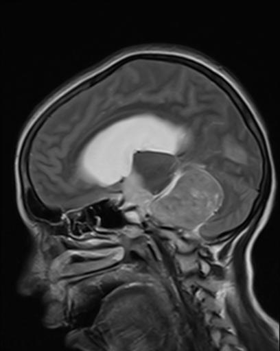
File:Acoustic schwannoma (Radiopaedia 20391-20296 Sagittal T2 11).jpg
Jump to navigation
Jump to search
Acoustic_schwannoma_(Radiopaedia_20391-20296_Sagittal_T2_11).jpg (408 × 512 pixels, file size: 18 KB, MIME type: image/jpeg)
Summary:
- Radiopaedia case ID: 20391
- Image ID: 2590016
- Image stack position: 11/66
- Plane projection: Sagittal
- Aux modality: T2
- Modality: MRI
- System: Central Nervous System
- Findings: There is a large well-defined extra-axial mass lesion seen in the left cerebellopontine (CP) angle region, extending into the left internal auditory canal with enlargement of the ipsilateral CP angle cistern. The lesion appears heterogeneously hyperintense on the T2W and FLAIR images, hypointense on T1W images and heterogeneously enhancing on contrast administration with enhancement of the left VII - VIII nerve complexes. There is widening of the left internal auditory canal. The lesion causes mass effect in the form of compressive buckling of the left cerebellar hemisphere, cerebellar peduncles, compressive rotation of the brain stem. The lesion causes compression and right lateral displacement of the fourth ventricle with moderate dilatation of both the lateral and the third ventricles.
- Published: 16th Nov 2012
- Source: https://radiopaedia.org/cases/acoustic-schwannoma-8
- Author: Vinay Shah
- Permission: http://creativecommons.org/licenses/by-nc-sa/3.0/
Licensing:
Attribution-NonCommercial-ShareAlike 3.0 Unported (CC BY-NC-SA 3.0)
File history
Click on a date/time to view the file as it appeared at that time.
| Date/Time | Thumbnail | Dimensions | User | Comment | |
|---|---|---|---|---|---|
| current | 02:03, 2 April 2021 |  | 408 × 512 (18 KB) | Fæ (talk | contribs) | Radiopaedia project rID:20391 (batch #489-80 E11) |
You cannot overwrite this file.
File usage
The following page uses this file:
