File:Acoustic schwannoma (Radiopaedia 39170-41387 Axial FIESTA 106).png
Jump to navigation
Jump to search
No higher resolution available.
Acoustic_schwannoma_(Radiopaedia_39170-41387_Axial_FIESTA_106).png (512 × 512 pixels, file size: 247 KB, MIME type: image/png)
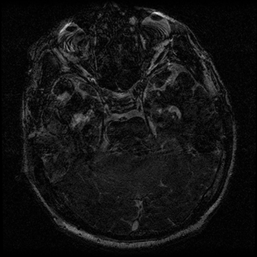
Summary:
- Radiopaedia case ID: 39170
- Image ID: 15401112
- Image stack position: 106/112
- Plane projection: Axial
- Aux modality: FIESTA
- Study description: MRI Brain - Posterior fossa
- Study findings: Extra-axial mass in the right cerebellopontine angle. The lesion has low to intermediate signal on T2 weighted images and intermediate T1 signal. There is homogeneous enhancement post administration of contrast. There is a possibility of peripheral nonenhancing cystic elements around the more solid enhancing lesion. The lesion bulges into the porus acousticus which appears mildly expanded. On the high resolution FIESTA images the nerves in the internal acoustic meatus can be seen separately to the mass. The superior aspect of the lesion abuts the undersurface of the tentorium with a possible dural tail in this region. Laterally the lesion abuts the petrous ridge, extending to the region of the jugular foramen. There is moderate edema in the adjacent cerebellar hemisphere and there is moderate mass effect on the right middle cerebral peduncle. Compressed vessels are seen around the lesion. There is distortion of the fourth ventricle and mild hydrocephalus seen involving the lateral and third ventricles. Conclusion: Right cerebellopontine angle mass with moderate local mass effect. The differential diagnosis is between meningioma and vestibular schwannoma, with meningioma slightly favored on the imaging appearance but both lesions remain in the differential diagnosis.
- Modality: MRI
- System: Oncology
- Findings: Right cerebellopontine angle mass is noted causing mass effect on the adjacent right cerebellum and slight distortion of the fourth ventricle but does not occlude it. No significant ventricular dilation. Remainder brain is unremarkable. Further investigation with MRI is recommended.
- Published: 24th Aug 2015
- Source: https://radiopaedia.org/cases/acoustic-schwannoma-21
- Author: Bruno Di Muzio
- Permission: http://creativecommons.org/licenses/by-nc-sa/3.0/
Licensing:
Attribution-NonCommercial-ShareAlike 3.0 Unported (CC BY-NC-SA 3.0)
File history
Click on a date/time to view the file as it appeared at that time.
| Date/Time | Thumbnail | Dimensions | User | Comment | |
|---|---|---|---|---|---|
| current | 06:21, 2 April 2021 | | 512 × 512 (247 KB) | Fæ (talk | contribs) | Radiopaedia project rID:39170 (batch #494-204 E106) |
You cannot overwrite this file.
File usage
The following page uses this file:
.png&oldid=114741)