File:Acquired cerebellar tonsillar ectopia (Radiopaedia 39950-42423 Axial FLAIR 16).jpg
Jump to navigation
Jump to search
No higher resolution available.
Acquired_cerebellar_tonsillar_ectopia_(Radiopaedia_39950-42423_Axial_FLAIR_16).jpg (392 × 512 pixels, file size: 22 KB, MIME type: image/jpeg)
Summary:
- Radiopaedia case ID: 39950
- Image ID: 16307490
- Image stack position: 16/18
- Plane projection: Axial
- Aux modality: FLAIR
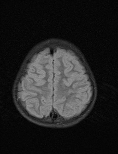
- Study findings: Normal sized posterior fossa. Engorgement of the venous sinuses and cortical veins as a result of negative CSF pressure. Herniated cerebellar tonsils through the foramen magnum by around 3 cm. Right cerebellopontine angle (CPA) lesion with fluid instensity on T2WI indenting the pons and right cerebellar hemisphere. The pons is displaced to the left side and the left CPA cistern is obliterated. On FLAIR, the lesion's fluid signal is suppressed, confirming its CSF content. The lesion shows a bright signal on DWI, but high ADC values denote no diffusion restriction, ruling out epidermoid.
- Modality: MRI
- System: Paediatrics
- Findings: Plain radiography showing a lumboperitoneal shunt catheter. One free end of the shunt catheter is seen in the peritoneal cavity, with the other end seen in place in the spinal canal at the upper lumbar spine. Lower lumbar and sacral spina bifida, along with pelvic bony deformity.
- Published: 15th Oct 2015
- Source: https://radiopaedia.org/cases/acquired-cerebellar-tonsillar-ectopia
- Author: Mahmoud Yacout Alabd
- Permission: http://creativecommons.org/licenses/by-nc-sa/3.0/
Licensing:
Attribution-NonCommercial-ShareAlike 3.0 Unported (CC BY-NC-SA 3.0)
File history
Click on a date/time to view the file as it appeared at that time.
| Date/Time | Thumbnail | Dimensions | User | Comment | |
|---|---|---|---|---|---|
| current | 19:03, 2 April 2021 | | 392 × 512 (22 KB) | Fæ (talk | contribs) | Radiopaedia project rID:39950 (batch #523-53 C16) |
You cannot overwrite this file.
File usage
There are no pages that use this file.
.jpg&oldid=120901)