File:Acquired cerebellar tonsillar ectopia (Radiopaedia 39950-42424 Sagittal T1 6).jpg
Jump to navigation
Jump to search
No higher resolution available.
Acquired_cerebellar_tonsillar_ectopia_(Radiopaedia_39950-42424_Sagittal_T1_6).jpg (224 × 256 pixels, file size: 11 KB, MIME type: image/jpeg)
Summary:
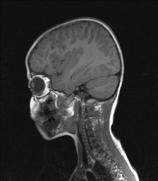
- Radiopaedia case ID: 39950
- Image ID: 16350034
- Image stack position: 6/23
- Plane projection: Sagittal
- Aux modality: T1
- Study description: MR study done 3 years prior
- Study findings: Normal location of the cerebellar tonsils. Normal size of the posterior fossa. The right CPA lesion is not visible.
- Modality: MRI
- System: Paediatrics
- Findings: Plain radiography showing a lumboperitoneal shunt catheter. One free end of the shunt catheter is seen in the peritoneal cavity, with the other end seen in place in the spinal canal at the upper lumbar spine. Lower lumbar and sacral spina bifida, along with pelvic bony deformity.
- Published: 15th Oct 2015
- Source: https://radiopaedia.org/cases/acquired-cerebellar-tonsillar-ectopia
- Author: Mahmoud Yacout Alabd
- Permission: http://creativecommons.org/licenses/by-nc-sa/3.0/
Licensing:
Attribution-NonCommercial-ShareAlike 3.0 Unported (CC BY-NC-SA 3.0)
File history
Click on a date/time to view the file as it appeared at that time.
| Date/Time | Thumbnail | Dimensions | User | Comment | |
|---|---|---|---|---|---|
| current | 19:05, 2 April 2021 | | 224 × 256 (11 KB) | Fæ (talk | contribs) | Radiopaedia project rID:39950 (batch #523-6 A6) |
You cannot overwrite this file.
File usage
There are no pages that use this file.
.jpg&oldid=120987)