File:Acquired hepatocerebral degeneration (Radiopaedia 41822-44802 Axial T1 27).jpg
Jump to navigation
Jump to search
Size of this preview: 540 × 599 pixels. Other resolutions: 216 × 240 pixels | 627 × 696 pixels.
Original file (627 × 696 pixels, file size: 27 KB, MIME type: image/jpeg)
Summary:
- Radiopaedia case ID: 41822
- Image ID: 18314922
- Image stack position: 27/27
- Plane projection: Axial
- Aux modality: T1
- Study description: MRI Brain
- Modality: MRI
- System: Hepatobiliary
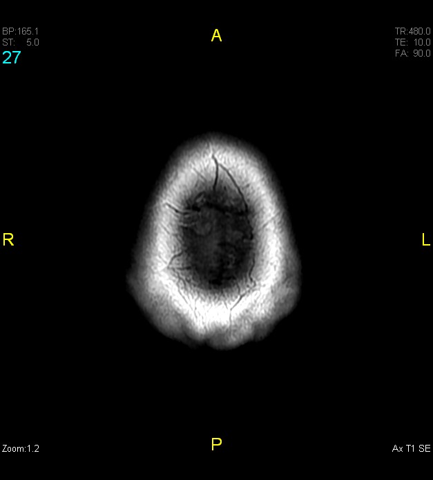
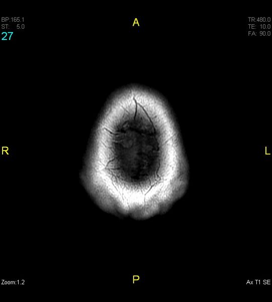
- Findings: Symmetric T1 hyperintense signals are seen arising from the globi pallidi, ventrolateral thalami, and substantia nigra of the midbrain. There is sparing of the cerebellum, red nucleus, and putamen. No features of midbrain atrophy. Age proportionate cerebral atrophic changes.
- Published: 22nd Dec 2015
- Source: https://radiopaedia.org/cases/acquired-hepatocerebral-degeneration
- Author: Varun Babu
- Permission: http://creativecommons.org/licenses/by-nc-sa/3.0/
Licensing:
Attribution-NonCommercial-ShareAlike 3.0 Unported (CC BY-NC-SA 3.0)
File history
Click on a date/time to view the file as it appeared at that time.
| Date/Time | Thumbnail | Dimensions | User | Comment | |
|---|---|---|---|---|---|
| current | 23:21, 2 April 2021 | | 627 × 696 (27 KB) | Fæ (talk | contribs) | Radiopaedia project rID:41822 (batch #530-27 A27) |
You cannot overwrite this file.
File usage
There are no pages that use this file.
.jpg&oldid=122297)