File:Acquired tracheo-esophageal fistula (Radiopaedia 51356-57103 Axial C+ arterial phase 5).jpg
Jump to navigation
Jump to search

Size of this preview: 800 × 467 pixels. Other resolutions: 320 × 187 pixels | 640 × 373 pixels | 1,024 × 597 pixels | 1,339 × 781 pixels.
Original file (1,339 × 781 pixels, file size: 73 KB, MIME type: image/jpeg)
Summary:
- Radiopaedia case ID: 51356
- Image ID: 28705948
- Image stack position: 5/49
- Plane projection: Axial
- Aux modality: C+ arterial phase
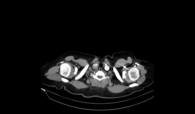
- Study findings: Necrotic nodal mass in the paratracheal chain extending inferiorly to the level of the carina. There is evidence of abnormal communication between the esophagus and trachea just above the level of the carina. A cavitary lesion in the apical segment of the right upper lobe and a soft tissue density nodule with some spiculated outlines in the posterior segment of the right upper lobe. Aspirated contrast material is noted in bilateral lung lower lobes mostly on the right side.
- Modality: CT
- System: Chest
- Findings: Upper GI barium study shows complete obliteration of the proximal esophagus with abnormal opacification of the right lower lobe bronchial tree.
- Published: 15th Apr 2017
- Source: https://radiopaedia.org/cases/acquired-tracheo-oesophageal-fistula-1
- Author: Hidayatullah Hamidi
- Permission: http://creativecommons.org/licenses/by-nc-sa/3.0/
Licensing:
Attribution-NonCommercial-ShareAlike 3.0 Unported (CC BY-NC-SA 3.0)
File history
Click on a date/time to view the file as it appeared at that time.
| Date/Time | Thumbnail | Dimensions | User | Comment | |
|---|---|---|---|---|---|
| current | 00:40, 3 April 2021 | | 1,339 × 781 (73 KB) | Fæ (talk | contribs) | Radiopaedia project rID:51356 (batch #537-5 A5) |
You cannot overwrite this file.
File usage
There are no pages that use this file.
.jpg&oldid=122600)