File:Acromial fracture (Radiopaedia 30094-30683 Axial view 1).jpg
Jump to navigation
Jump to search
Size of this preview: 476 × 599 pixels. Other resolutions: 191 × 240 pixels | 381 × 480 pixels | 610 × 768 pixels | 814 × 1,024 pixels | 2,400 × 3,020 pixels.
Original file (2,400 × 3,020 pixels, file size: 255 KB, MIME type: image/jpeg)
Summary:
- Radiopaedia case ID: 30094
- Image ID: 7644308
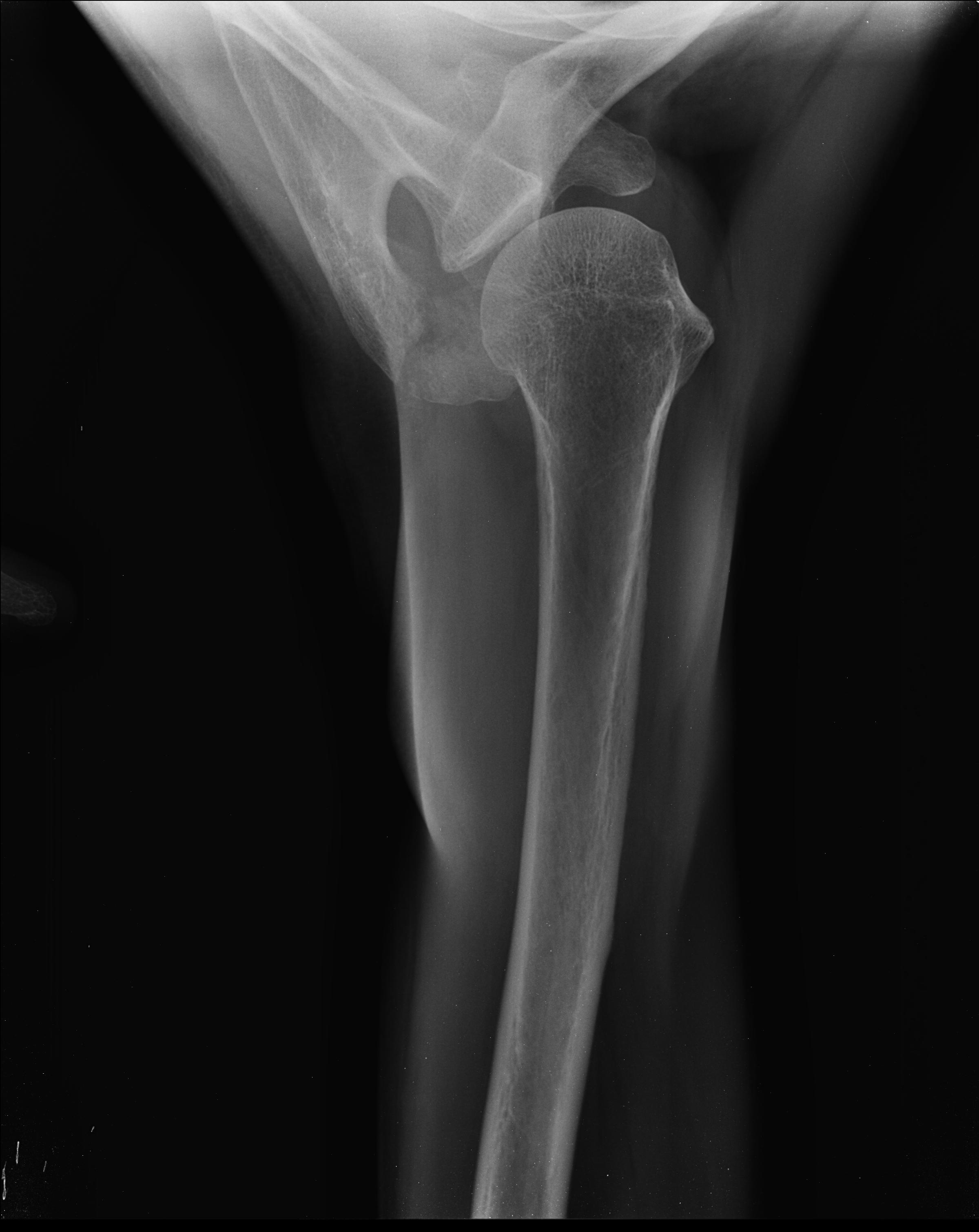
- Plane projection: Axial view
- Study description: Ultrasound findings favored acromial fracture over less likely os acromiale. Shoulder axial view radiograph was ordered to differentiate them.
- Study findings: Fracture line through the acromion process of the scapula. If corticated margins are noted on both sided of lucency, it favors os acromiale.
- Modality: X-ray
- System: Trauma
- Findings: Intact and normal echo pattern of the rotator cuff. No bursal effusion. No glenohumeral effusion. Acromion shows cortical breach, fragmentation.
- Published: 18th Jul 2014
- Source: https://radiopaedia.org/cases/acromial-fracture-1
- Author: Maulik S Patel
- Permission: http://creativecommons.org/licenses/by-nc-sa/3.0/
Licensing:
Attribution-NonCommercial-ShareAlike 3.0 Unported (CC BY-NC-SA 3.0)
File history
Click on a date/time to view the file as it appeared at that time.
| Date/Time | Thumbnail | Dimensions | User | Comment | |
|---|---|---|---|---|---|
| current | 01:41, 3 April 2021 | | 2,400 × 3,020 (255 KB) | Fæ (talk | contribs) | Radiopaedia project rID:30094 (batch #552-1 A1) |
You cannot overwrite this file.
File usage
There are no pages that use this file.
.jpg&oldid=123230)