File:Acromioclavicular injury - Rockwood type V (Radiopaedia 85749-101842 Coronal bone window 30).jpg
Jump to navigation
Jump to search
Size of this preview: 800 × 373 pixels. Other resolutions: 320 × 149 pixels | 640 × 298 pixels | 1,098 × 512 pixels.
Original file (1,098 × 512 pixels, file size: 182 KB, MIME type: image/jpeg)
Summary:
- Radiopaedia case ID: 85749
- Image ID: 54232351
- Image stack position: 30/43
- Plane projection: Coronal
- Aux modality: bone window
- Study description: CT Left Shoulder
- Study findings: CT confirms the findings of acromioclavicular dislocation.
- Modality: CT
- System: Musculoskeletal
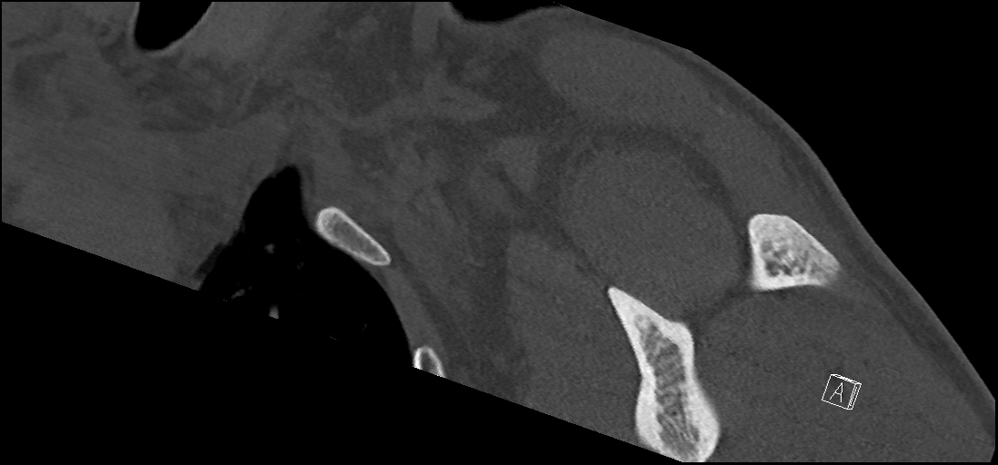
- Findings: There is dislocation of the left acromioclavicular joint (ACJ) with loss of normal alignment of the inferior surfaces of clavicle and acromion. The inferior border of the clavicle is elevated beyond the superior border of the acromion in the dedicated AC joint view. There is increased coracoclavicular distance on the affected side. It is 30.5 mm on the symptomatic left compared to 11.5mm on right. (Normal coracoclavicular distance is 11 - 13mm). So it is more than double the normal and indirect evidence of coracoclavicular ligament injury. Hence suggestive of Rockwood type V injury. Comparison is done with the contralateral acromioclavicular joint, which is in normal alignment. The glenohumeral joint is in normal alignment Head, neck and proximal shaft of the left humerus are normal.
- Published: 19th Jan 2021
- Source: https://radiopaedia.org/cases/acromioclavicular-injury-rockwood-type-v
- Author: Saneej Kanhirat
- Permission: http://creativecommons.org/licenses/by-nc-sa/3.0/
Licensing:
Attribution-NonCommercial-ShareAlike 3.0 Unported (CC BY-NC-SA 3.0)
File history
Click on a date/time to view the file as it appeared at that time.
| Date/Time | Thumbnail | Dimensions | User | Comment | |
|---|---|---|---|---|---|
| current | 02:53, 3 April 2021 | | 1,098 × 512 (182 KB) | Fæ (talk | contribs) | Radiopaedia project rID:85749 (batch #567-30 A30) |
You cannot overwrite this file.
File usage
There are no pages that use this file.
.jpg&oldid=124284)