File:Actinomycosis - pulmonary manifestations on chest radiographs (Radiopaedia 38716-40877 Lateral 1).jpg
Jump to navigation
Jump to search

Size of this preview: 484 × 600 pixels. Other resolutions: 194 × 240 pixels | 387 × 480 pixels | 620 × 768 pixels | 826 × 1,024 pixels | 1,624 × 2,012 pixels.
Original file (1,624 × 2,012 pixels, file size: 1.14 MB, MIME type: image/jpeg)
Summary:
- Radiopaedia case ID: 38716
- Image ID: 14956974
- Plane projection: Lateral
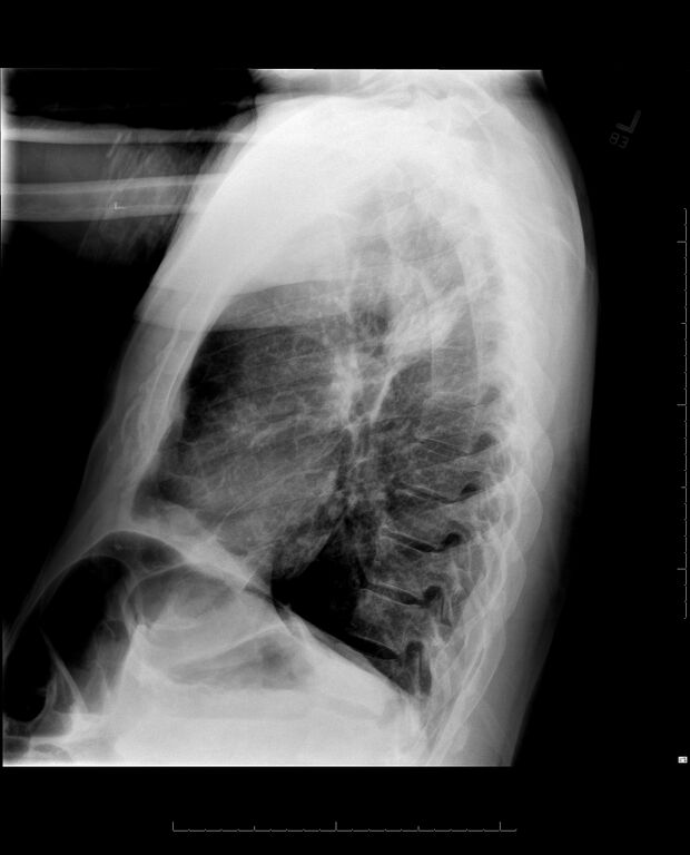
- Study description: 8 years later the chest X ray shows
- Study findings: Right upper lobe findings are similar. Grossly no change.
- Modality: X-ray
- System: Chest
- Findings: Frontal and lateral view of the chest demonstrate chronic interstitial lung markings, which are actually more prominent in the right upper lobe and right lower lobe suggestive of scarring. This is stable for 10 years. There is interposition of colon between liver and right hemidiaphragm.
- Published: 30th Jul 2015
- Source: https://radiopaedia.org/cases/actinomycosis-pulmonary-manifestations-on-chest-radiographs-1
- Author: Jayanth Keshavamurthy
- Permission: http://creativecommons.org/licenses/by-nc-sa/3.0/
Licensing:
Attribution-NonCommercial-ShareAlike 3.0 Unported (CC BY-NC-SA 3.0)
| This file is ineligible for copyright and therefore in the public domain, because it is a technical image created as part of a standard medical diagnostic procedure. No creative element rising above the threshold of originality was involved in its production.
|
 |
File history
Click on a date/time to view the file as it appeared at that time.
| Date/Time | Thumbnail | Dimensions | User | Comment | |
|---|---|---|---|---|---|
| current | 11:06, 3 April 2021 | | 1,624 × 2,012 (1.14 MB) | Fæ (talk | contribs) | Radiopaedia project rID:38716 (batch #623-2 B1) |
You cannot overwrite this file.
File usage
There are no pages that use this file.
.jpg&oldid=9754198)