File:Active bleeding from duodenal ulcer with embolization (Radiopaedia 34216-35481 Axial C+ arterial phase 30).png
Jump to navigation
Jump to search
No higher resolution available.
Active_bleeding_from_duodenal_ulcer_with_embolization_(Radiopaedia_34216-35481_Axial_C+_arterial_phase_30).png (512 × 512 pixels, file size: 161 KB, MIME type: image/png)
Summary:
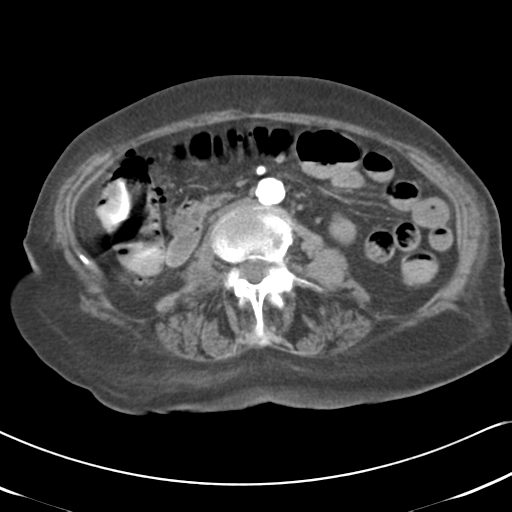
- Radiopaedia case ID: 34216
- Image ID: 10960881
- Image stack position: 30/74
- Plane projection: Axial
- Aux modality: C+ arterial phase
- Modality: CT
- System: Gastrointestinal
- Findings: On the arterial and portal venous phase images there is a 1 cm region of vivid enhancement in the first part of duodenum, that was not present on the non-contrast images. This region receives supply from the gastroduodenal artery. No intraluminal contrast extravasation is seen elsewhere in the bowel. Gallstones incidentally noted. The liver, spleen, pancreas and right kidney are unremarkable. The left kidney is mildly atrophic with regions of cortical scarring. A small left and tiny right pleural effusion is noted. Conclusion: The site of active hemorrhage is localized to the proximal duodenum.
- Published: 10th Feb 2015
- Source: https://radiopaedia.org/cases/active-bleeding-from-duodenal-ulcer-with-embolisation
- Author: RMH Core Conditions
- Permission: http://creativecommons.org/licenses/by-nc-sa/3.0/
Licensing:
Attribution-NonCommercial-ShareAlike 3.0 Unported (CC BY-NC-SA 3.0)
File history
Click on a date/time to view the file as it appeared at that time.
| Date/Time | Thumbnail | Dimensions | User | Comment | |
|---|---|---|---|---|---|
| current | 11:49, 3 April 2021 | | 512 × 512 (161 KB) | Fæ (talk | contribs) | Radiopaedia project rID:34216 (batch #625-105 B30) |
You cannot overwrite this file.
File usage
The following page uses this file:
.png&oldid=104870)