File:Active colonic bleed on CT (Radiopaedia 49765-55025 Coronal delay MIP 30).jpg
Jump to navigation
Jump to search
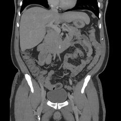
Size of this preview: 600 × 600 pixels. Other resolutions: 240 × 240 pixels | 608 × 608 pixels.
Original file (608 × 608 pixels, file size: 74 KB, MIME type: image/jpeg)
Summary:
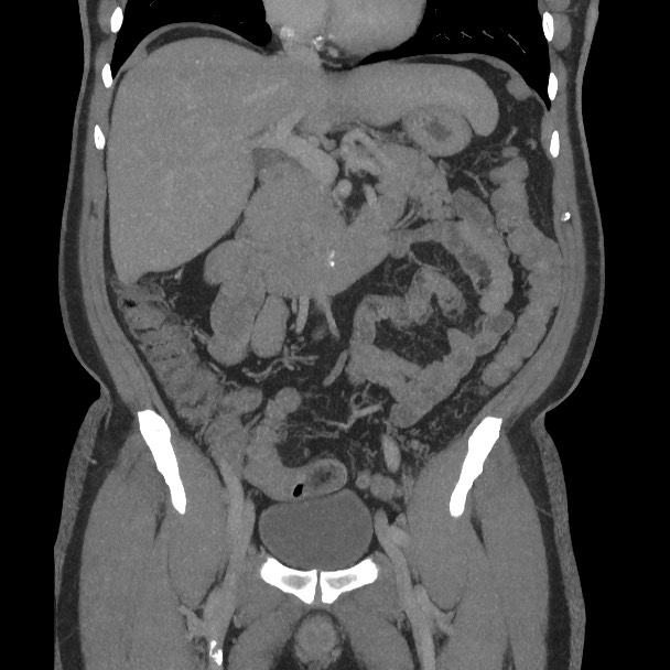
- Radiopaedia case ID: 49765
- Image ID: 26796660
- Image stack position: 30/68
- Plane projection: Coronal
- Aux modality: delay MIP
- Modality: CT
- System: Gastrointestinal
- Findings: Active extravasation of contrast into the large bowel lumen at the junction of the descending colon and sigmoid colon is seen on the arterial phase study with further pooling of contrast on the delayed phase. There is no mass lesion at this site to suggest bleeding from a colonic malignancy, nor is there any surrounding inflammatory change to suggest diverticulitis as a cause.
- Published: 15th Feb 2017
- Source: https://radiopaedia.org/cases/active-colonic-bleed-on-ct
- Author: Andrew Dixon
- Permission: http://creativecommons.org/licenses/by-nc-sa/3.0/
Licensing:
Attribution-NonCommercial-ShareAlike 3.0 Unported (CC BY-NC-SA 3.0)
File history
Click on a date/time to view the file as it appeared at that time.
| Date/Time | Thumbnail | Dimensions | User | Comment | |
|---|---|---|---|---|---|
| current | 13:43, 3 April 2021 | | 608 × 608 (74 KB) | Fæ (talk | contribs) | Radiopaedia project rID:49765 (batch #627-445 F30) |
You cannot overwrite this file.
File usage
The following page uses this file:
.jpg&oldid=106189)