File:Active diverticular hemorrhage (Radiopaedia 39415-41725 Axial C+ portal venous phase 16).png
Jump to navigation
Jump to search
No higher resolution available.
Active_diverticular_hemorrhage_(Radiopaedia_39415-41725_Axial_C+_portal_venous_phase_16).png (512 × 512 pixels, file size: 194 KB, MIME type: image/png)
Summary:
- Radiopaedia case ID: 39415
- Image ID: 15672927
- Image stack position: 16/83
- Plane projection: Axial
- Aux modality: C+ portal venous phase
- Modality: CT
- System: Gastrointestinal
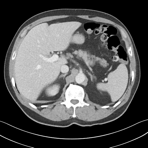
- Findings: Extravasation of contrast within the mid sigmoid colon, with pooling on the portal venous phase. Extensive colonic diverticular disease. The remainder of the colon is unremarkable aside from some fecal colonic loading. Incidental duodenal diverticulum. Liver, spleen, adrenal glands, pancreas are normal. Cholelithiasis. Multiple bilateral non-obstructive renal stones. Multiple simple renal cortical cysts. Prostatomegaly. Left ureterocoele or bladder diverticulum with calcification, which may be in the wall or may represent a dependent stone. No free fluid or free gas.
- Published: 3rd Sep 2015
- Source: https://radiopaedia.org/cases/active-diverticular-haemorrhage
- Author: Henry Knipe
- Permission: http://creativecommons.org/licenses/by-nc-sa/3.0/
Licensing:
Attribution-NonCommercial-ShareAlike 3.0 Unported (CC BY-NC-SA 3.0)
File history
Click on a date/time to view the file as it appeared at that time.
| Date/Time | Thumbnail | Dimensions | User | Comment | |
|---|---|---|---|---|---|
| current | 16:15, 3 April 2021 | | 512 × 512 (194 KB) | Fæ (talk | contribs) | Radiopaedia project rID:39415 (batch #630-182 C16) |
You cannot overwrite this file.
File usage
The following page uses this file:
.png&oldid=107831)