File:Active renal extravasation with large subcapsular and retroperitoneal hemorrhage (Radiopaedia 60975-68796 Axial C+ arterial phase 136).jpg
Jump to navigation
Jump to search
No higher resolution available.
Active_renal_extravasation_with_large_subcapsular_and_retroperitoneal_hemorrhage_(Radiopaedia_60975-68796_Axial_C+_arterial_phase_136).jpg (512 × 512 pixels, file size: 64 KB, MIME type: image/jpeg)
Summary:
| Description |
|
| Date | 11 Jun 2018 |
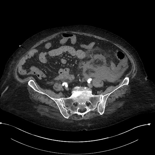
| Source | Active renal extravasation with large subcapsular and retroperitoneal hemorrhage |
| Author | Michael P Hartung |
| Permission (Permission-reusing-text) |
http://creativecommons.org/licenses/by-nc-sa/3.0/ |
Licensing:
Attribution-NonCommercial-ShareAlike 3.0 Unported (CC BY-NC-SA 3.0)
| This file is ineligible for copyright and therefore in the public domain, because it is a technical image created as part of a standard medical diagnostic procedure. No creative element rising above the threshold of originality was involved in its production.
|
 |
File history
Click on a date/time to view the file as it appeared at that time.
| Date/Time | Thumbnail | Dimensions | User | Comment | |
|---|---|---|---|---|---|
| current | 20:18, 3 April 2021 | | 512 × 512 (64 KB) | Fæ (talk | contribs) | Radiopaedia project rID:60975 (batch #636-136 A136) |
You cannot overwrite this file.
File usage
The following page uses this file:
.jpg&oldid=9702469)