File:Active splenic bleeding (Radiopaedia 45152-49138 Coronal C+ portal venous phase 34).png
Jump to navigation
Jump to search
Size of this preview: 497 × 600 pixels. Other resolutions: 199 × 240 pixels | 512 × 618 pixels.
Original file (512 × 618 pixels, file size: 309 KB, MIME type: image/png)
Summary:
- Radiopaedia case ID: 45152
- Image ID: 22385997
- Image stack position: 34/60
- Plane projection: Coronal
- Aux modality: C+ portal venous phase
- Study description: CT Abdomen and pelvis
- Modality: CT
- System: Hepatobiliary
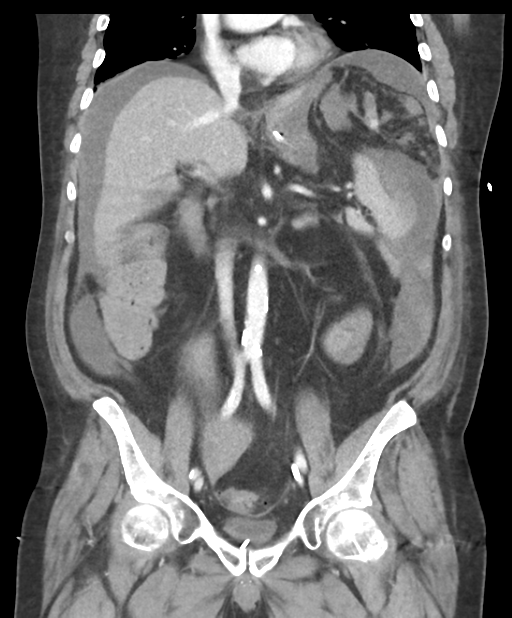
- Findings: The amount of free fluid in the peritoneal cavity is overall stable compared to the previous imaging, although there is now active hemorrhage on the lateral aspect of the spleen characterized by contrast extravasation within a subcapsular hematoma. The hemorrhage tracks down to the pelvis and mix with the ascites elevating its average attenuation. There is small volume fluid extending through a right lateral inferior abdominal wall hernia. Multiple colonic diverticula. Bowel is otherwise unremarkable. No free gas. The liver, pancreas, and adrenal glands are unremarkable. Multiple small calcified gallstones. Kidneys have reduced dimensions. No hydronephrosis. There is no lymph node enlargement. No suspicious bone lesions are seen. Small left side pleural effusion. Bilateral gynecomastia. Ill-defined fat stranding and small subcutaneous hematoma in the anterior abdominal wall are probably related to previous catheter manipulations.
- Published: 29th May 2016
- Source: https://radiopaedia.org/cases/active-splenic-bleeding
- Author: Bruno Di Muzio
- Permission: http://creativecommons.org/licenses/by-nc-sa/3.0/
Licensing:
Attribution-NonCommercial-ShareAlike 3.0 Unported (CC BY-NC-SA 3.0)
File history
Click on a date/time to view the file as it appeared at that time.
| Date/Time | Thumbnail | Dimensions | User | Comment | |
|---|---|---|---|---|---|
| current | 00:17, 4 April 2021 | | 512 × 618 (309 KB) | Fæ (talk | contribs) | Radiopaedia project rID:45152 (batch #640-134 B34) |
You cannot overwrite this file.
File usage
The following page uses this file:
.png&oldid=113142)