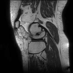
File:Acute-on-chronic transient lateral patellar dislocation with trochlear dysplasia (Radiopaedia 84099-99349 Sagittal PD 126).jpg
Jump to navigation
Jump to search
No higher resolution available.
Acute-on-chronic_transient_lateral_patellar_dislocation_with_trochlear_dysplasia_(Radiopaedia_84099-99349_Sagittal_PD_126).jpg (256 × 256 pixels, file size: 16 KB, MIME type: image/jpeg)
Summary:
| Description |
|
| Date | Published: 27th Mar 2021 |
| Source | https://radiopaedia.org/cases/acute-on-chronic-transient-lateral-patellar-dislocation-with-trochlear-dysplasia |
| Author | Henry Knipe |
| Permission (Permission-reusing-text) |
http://creativecommons.org/licenses/by-nc-sa/3.0/ |
Licensing:
Attribution-NonCommercial-ShareAlike 3.0 Unported (CC BY-NC-SA 3.0)
File history
Click on a date/time to view the file as it appeared at that time.
| Date/Time | Thumbnail | Dimensions | User | Comment | |
|---|---|---|---|---|---|
| current | 08:32, 16 April 2021 | | 256 × 256 (16 KB) | Fæ (talk | contribs) | Radiopaedia project rID:84099 (batch #967-202 C126) |
You cannot overwrite this file.
File usage
The following page uses this file:
.jpg&oldid=147250)