File:Acute A3 occlusion with ACA ischemic penumbra (CT perfusion) (Radiopaedia 72036-82528 MTT 10).jpg
Jump to navigation
Jump to search
Size of this preview: 600 × 600 pixels. Other resolutions: 240 × 240 pixels | 480 × 480 pixels | 768 × 768 pixels | 1,024 × 1,024 pixels | 2,324 × 2,324 pixels.
Original file (2,324 × 2,324 pixels, file size: 992 KB, MIME type: image/jpeg)
Summary:
- Radiopaedia case ID: 72036
- Image ID: 51709669
- Image stack position: 10/31
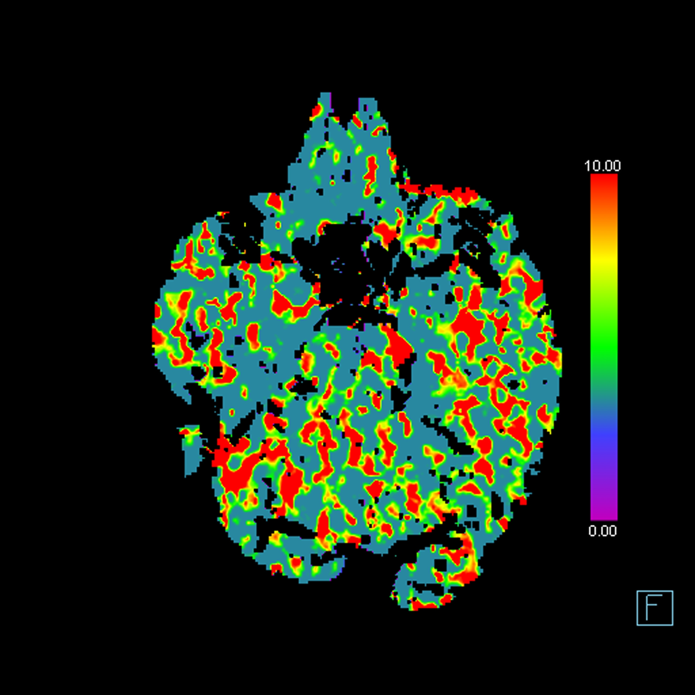
- Aux modality: MTT
- Study description: CT perfusion
- Study findings: Increased time parameters in the posterior medial left cerebral hemisphere indicating distal left ACA penumbra. No evidence of core infarct.
- Modality: CT
- System: Interventional
- Findings: No hemorrhage, surface collection, mass effect or midline shift. Grey-white matter differentiation is preserved. Small left A3 dense vessel sign on the thin data set. The ventricles and basal cisterns are symmetric and normal for age.
- Published: 5th Nov 2019
- Source: https://radiopaedia.org/cases/acute-a3-occlusion-with-aca-ischaemic-penumbra-ct-perfusion
- Author: Craig Hacking
- Permission: http://creativecommons.org/licenses/by-nc-sa/3.0/
Licensing:
Attribution-NonCommercial-ShareAlike 3.0 Unported (CC BY-NC-SA 3.0)
File history
Click on a date/time to view the file as it appeared at that time.
| Date/Time | Thumbnail | Dimensions | User | Comment | |
|---|---|---|---|---|---|
| current | 02:05, 4 April 2021 | | 2,324 × 2,324 (992 KB) | Fæ (talk | contribs) | Radiopaedia project rID:72036 (batch #642-11 B10) |
You cannot overwrite this file.
File usage
There are no pages that use this file.
_(Radiopaedia_72036-82528_MTT_10).jpg&oldid=113682)