File:Acute Budd-Chiari syndrome (Radiopaedia 60858-68638 Axial C+ portal venous phase 102).jpg
Jump to navigation
Jump to search

Size of this preview: 800 × 589 pixels. Other resolutions: 320 × 236 pixels | 640 × 471 pixels | 1,024 × 754 pixels | 1,280 × 943 pixels | 1,798 × 1,324 pixels.
Original file (1,798 × 1,324 pixels, file size: 69 KB, MIME type: image/jpeg)
Summary:
- Radiopaedia case ID: 60858
- Image ID: 39596473
- Image stack position: 102/321
- Plane projection: Axial
- Aux modality: C+ portal venous phase
- Modality: CT
- System: Hepatobiliary
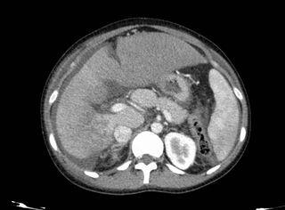
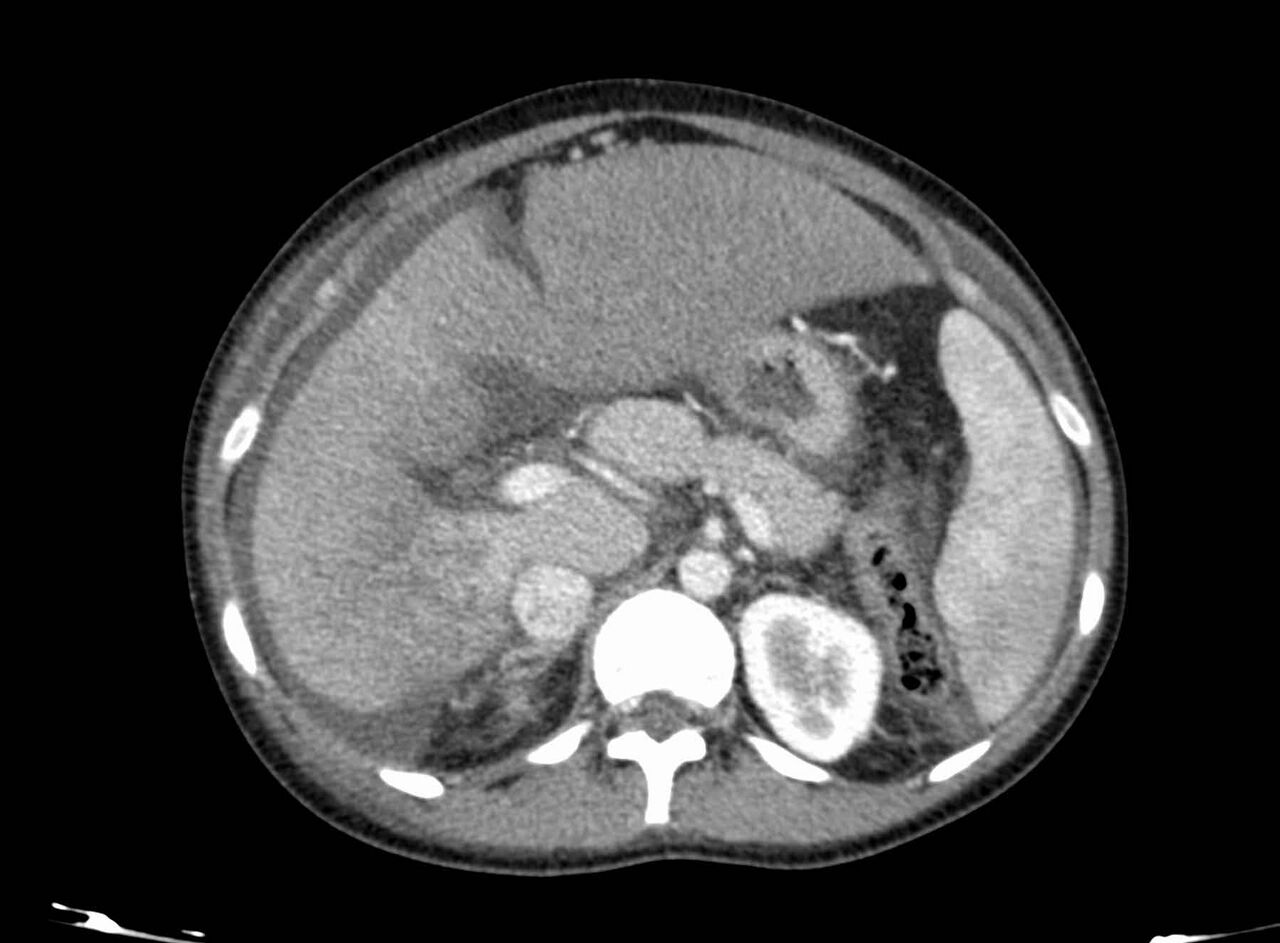
- Findings: Contrast-enhanced abdomino-pelvic CT scan in the portovenous phase shows a lack of enhancement of hepatic veins, filling defects in the IVC and ascites. There is a reduced and heterogeneous enhancement of liver parenchyma with sparing of the caudate lobe. This finding is seen due to separate venous drainage of this liver segment. Splenomegaly.
- Published: 5th Jun 2018
- Source: https://radiopaedia.org/cases/acute-budd-chiari-syndrome
- Author: Bita Abbasi
- Permission: http://creativecommons.org/licenses/by-nc-sa/3.0/
Licensing:
Attribution-NonCommercial-ShareAlike 3.0 Unported (CC BY-NC-SA 3.0)
File history
Click on a date/time to view the file as it appeared at that time.
| Date/Time | Thumbnail | Dimensions | User | Comment | |
|---|---|---|---|---|---|
| current | 01:18, 6 April 2021 | | 1,798 × 1,324 (69 KB) | Fæ (talk | contribs) | Radiopaedia project rID:60858 (batch #749-423 B102) |
You cannot overwrite this file.
File usage
The following page uses this file:
.jpg&oldid=73701)